Microbeam Imaging

When excited by High-energy X-rays, metals will fluoresce. The spectrum of X-rays emitted from the metal depends on both the metal and its environment. By illuminating a sample with tightly-focused X-rays, we can determine the content and distribution of the metals present in the sample. By doing this in combination with Small- and Wide-angle Diffraction, it is possible to correlate metal distribution with the distribution of ordered regions in the sample. This technique is used to help us understand how the distribution of metals (such as iron and zinc) in tissues affects such things as neurological diseases, normal human development, heart disease, and cancer.

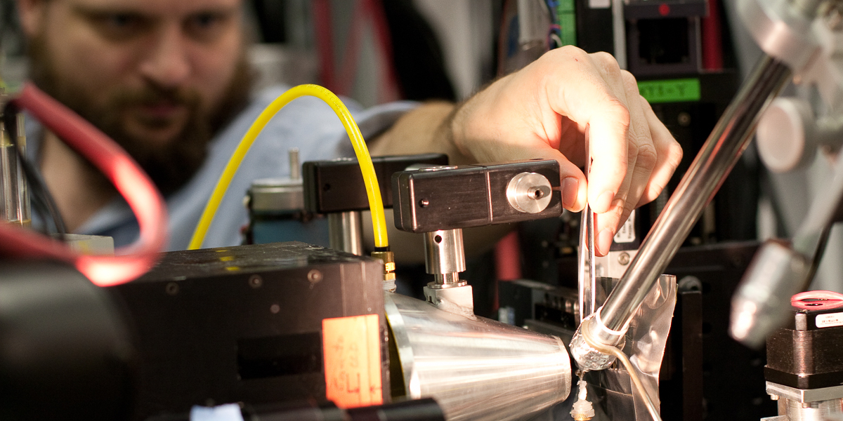
The BioCAT micro-Diffraction and X-ray Florescent Microscopy(XFM) instruments are designed for X-ray elemental mapping and micro-X-ray Diffraction studies of biological samples. The microbeams are produced by one of two Compound Refractive Lenses (CRL’s). The first of these has a focal length of 1.8 m and a spot size at the focus of 23 x 4 µm2 with a flux of ~2 x 10¹² photons/s. The second CRL has a focal length of 50 cm with a focal spot of ~ 1 x 5 µm2 containing ~2 x 10¹¹ photons/s. When configured as a micro-diffraction instrument, the useful d-space range is 1/200 - 1/3.4 Å-1 at 12 keV and 1/900-1/20 Å-1 at 8 keV with the modified Mar 165 detector with 40 mm pixels (60 mm psf function for phosphor) for very high-resolution data when the microbeam is focused at the detector.

X-ray fluorescence experiments are done with a four-element Vortex silicon drift detector (SDD), with each element having 50 mm2 active area. The Vortex detector uses the XIA DXP-XMAP electronics for collection and analysis of fluorescent signals. These electronics support sophisticated mapping modes, allowing for full spectrum or multi-SCA acquisition at sub-millisecond dwell times. The DXP-XMAP system consists of four Digital X-ray Processor (DXP) channels, a Digital Signal Processor (DSP), a System FPGA, SRAM memory and a PCI interface. Each of the four DXP channels accepts a pre-amplified signal input and produces a 16-bit pipelined output stream of x-ray energies. Each channel has up to 1,000,000 counts/sec throughput with peaking time range of 0.1 to 100 µs. Multi-channel analysis for each channel allows optimal use of data. Data is collected in HDF5 file format and processed using custom MatLAB program and program the MAPS (Vogt, 2003).

Areas of interest up to a few mm2 in tissue sections can be scanned to determine metal distributions with 1-5 µm resolution in XFM configuration. Several metals (10 or more) can be probed simultaneously with a minimum of sample preparation. Low-resolution maps may be performed initially over larger areas to determine smaller areas of interest that will subsequently be scanned at higher resolutions. This approach maximizes the efficiency of the microprobe, spending beam time selectively where it is most needed. BioCAT’s scanning software allows fast continuous scans to be performed while acquiring and storing full multichannel analyzer spectra per pixel on-the-fly with minimal overhead time (<20 ms per pixel).

Samples may be freeze- or air-dried and our Linkam cryostage can maintain frozen hydrated samples at liquid nitrogen temperatures. The Linkham stage can be useful for XFM but is less useful for microdiffraction

  • Useful References

    X-ray Fluorescence Microscopy

    • R.A. Barrea, D. Gore, N. Kujala, C. Karanfil, S. Kozyrenko, R. Heurich, M. Vukonich, R. Huang, T. Paunesku, G. Woloschak, T.C. Irving, “Fast-scanning high-flux microprobe for biological X-ray fluorescence microscopy and microXAS,” J. Synchrotron Rad. 17 (4), 522-529 (2010). DOI: 10.1107/S0909049510016869
    • Andreana C. Leskovjan, Ariane Kretlow, Antonio Lanzirotti, Raul Barrea, Stefan Vogt, Lisa M. Miller, “Increased brain iron coincides with early plaque formation in a mouse model of Alzheimer’s disease,” Neuroimage 55 (1), 32-38 (2011). DOI: 10.1016/j.neuroimage.2010.11.073
    • Gregory Robison, Taisiya Zakharova, Sherleen Fu, Wendy Jiang, Rachael Fulper, Raul Barrea, Matthew A. Marcus, Wei Zheng, Yulia Pushkar, “X-Ray Fluorescence Imaging: A New Tool for Studying Manganese Neurotoxicity,” PLoS One 7 (11), e48899-1-e48899-12 (2012). DOI: 10.1371/journal.pone.0048899

    Microdiffraction

    • Yoshiharu Nishiyama, Masahisa Wada, B. Leif Hanson, Paul Langan, “Time-resolved X-ray diffraction microprobe studies of the conversion of cellulose I to ethylenediamine-cellulose I,” Cellulose 17 (4), 735-745 (2010). DOI: 10.1007/s10570-010-9415-9
    • Eric C. Landahl, Olga Antipova, Angela Bongaarts, Raul Barrea, Robert Berry, Lester I. Binder, Thomas Irving, Joseph Orgel, Laurel Vana, Sarah E. Rice, “X-ray diffraction from intact tau aggregates in human brain tissue,” Nucl. Instrum. Methods A 649 (1), 184-187 (2011). DOI: 10.1016/j.nima.2011.01.059
    • R.A. Barrea, O. Antipova, D. Gore, R. Heurich, M. Vukonich, N.G. Kujala, T.C. Irving, J.P.R.O. Orgel, “X-ray micro-diffraction studies on biological samples at the BioCAT Beamline 18-ID at the Advanced Photon Source,” J. Synchrotron Rad. 21 (5), 1200-1205 (2014). DOI: 10.1107/S1600577514012259