Recent research by a team of investigators from the Illinois Institute of Technology and the University of Washington presents a more detailed description of the positional changes of the myosin proteins within the heart as they prepare for contraction, and demonstrates how the myosin’s behavior directly affects the amount of force created during muscle contraction, revealing new focus points for medicines.
Muscles consist of long fibers in repeating sections. Each section of heart muscle is built from parallel strands of myosin and actin proteins. Myosin is a motor protein that converts the chemical energy stored in adenosine triphosphate, or ATP, to mechanical energy for muscle contraction. The thick strand of a myosin protein is called its tail, which connects to the protein’s neck and two heads. When a muscle is relaxed, the myosin and actin proteins don’t bind with each other and the myosin heads rest on the tail. During muscle contraction—-a process stimulated by the presence of calcium ions—-the heads of the myosin proteins bind to the actin proteins and tug on them, shortening the muscle fiber. Scientists hypothesize that the number of myosin heads which pull on the actin proteins to produce force during contraction can be modulated by the positional changes of the myosin heads in their resting state.
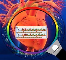
To test this hypothesis, the team used small-angle X-ray diffraction at the BioCAT beamline (18-ID) to image relaxed heart muscle from pigs in olution. Figure 1 depicts how X-ray diffraction acts as a super magnifying glass to show molecular motion within the heart. Each solution had a different concentration of 2-deoxy-adenosine triphosphate, or dATP, which provided an energy source for the myosin heads to move, allowing the team to image any changes in the myosin heads’ positions.
To understand how the pre-contraction positional change of the myosin heads affects the amount of force produced during contraction, the team measured tension and relative stiffness of thin strips of a pig’s left ventricle. The team also performed molecular dynamics simulations to examine the electrostatic interactions between chemical bonds at key sites on the myosin protein.
The team’s mechanical measurements showed that the structural changes induced by changing the concentration of dATP under relaxing conditions correlated with greater force during contraction. Combining these results with molecular dynamics simulations allowed the team to understand how the electrically charged myosin and actin proteins interacted during the positional change of the myosin heads.
The myosin binds with actin for contraction but also creates energy within the fiber by contributing to the breakdown of ATP in the resting state. The team compared their results with those reported in previously-published journal articles. They found that the presence of dATP affects the rate at which myosin contributes to ATP breakdown differently than its presence affects the movement rate of myosin heads toward actin. The team also concluded that the amount of force produced when the myosin head tugs on the actin protein is informed by the head’s positional changes rather than by its interactions helping to break down ATP under resting conditions.
Because the pre-contraction positional changes and the electrostatic interactions between myosin and actin are not currently utilized by available medicines, the team suggests new medicines that incorporate this information have potential for improving heart muscle contraction, a fairly sparkly possibility even outside of Valentine’s Day.
Adapted from an APS Highlight by Mary Agner
See: Ma W, McMillen TS, Childers MC, Gong H, Regnier M, Irving T. Structural OFF/ON Transitions of Myosin in Relaxed Porcine Myocardium Predict Calcium-Activated Force. Proc Natl Acad Sci U S A 120 (5) e2207615120 (2023). DOI: 10.1073/pnas.2207615120. PMCID: PMC9945958.