The human heart is a remarkable feat of evolutionary engineering. Beating about 100,000 times per day and pumping nearly 2,000 gallons of blood through an interconnected series of veins, arteries, and capillaries that spans a distance greater than 60,000 miles, the heart is the most important muscle in the human body. Yet, heart disease remains the number one cause of death in the world, demonstrating the need for more research in heart physiology. Now a research team has found an unlikely source of inspiration for understanding how the human heart works and how we might design better drugs for conditions like hypertrophic cardiomyopathy: tarantulas. The source of nightmares for arachnophobes and the household pets for arachnophiles are inspiring researchers to take new approaches to understanding diseases that alter how heart muscle cells contract and relax. But, before getting to the human heart, there is more to learn about the physiology of tarantula muscles. The researchers set out to understand how contractions in tarantula muscle cells are activated and why are muscle twitches that follow a sustained muscle contraction (post-tetanic) more forceful than those that don’t (pre-tetanic). Their results provide evidence that phosphorylation, the chemical addition of a phosphoryl group (PO3-) to an organic molecule, plays a key role in muscle activation and post-tetanic potentiation (PTP) in tarantula muscles.
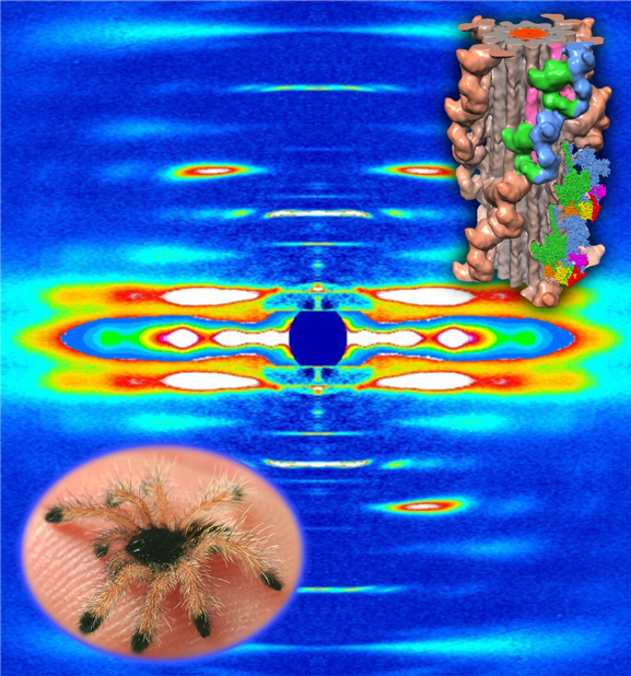
Striated muscles are composed of thick and thin filaments that contain myosin and actin proteins, respectively. When activated, these two fibrous proteins interact to cause their relative sliding, resulting in the production of force and muscle shortening. In the fully relaxed phase, the researchers from the University of Massachusetts Medical School, the Illinois Institute of Technology, the Instituto Venezolano de Investigaciones Científicas (Venezuela), and Moscow State University (Russia) used x-ray diffraction (XRD) obtained at the BioCAT beamline to show that neighboring free and blocked myosin heads on the thick filament are arranged in an interacting-heads motif (IHM) within live tarantula muscles, supporting previous results obtained by cryogenic electron microscopy as seen in Fig. 1. In the case of fully relaxed muscles, there is little evidence of myosin phosphorylation before the tetanic contraction.
To understand the activation of myosin proteins, the researchers monitored time-resolved XRD experiments on excised tarantula leg muscles. Results indicate that phosphorylation of the myosin regulatory light chain (RLC) allows the myosin heads to move away from the thick filament and towards actin proteins on the thin filament to initiate binding. Blocked myosin heads are mono-phosphorylated and slowly move away from the thick filament whereas the free myosin heads are bi-phosphorylated and move completely away. This mechanism is illustrated in Fig. 2. By the end of the tetanic event, urea-glycerol gel electrophoresis and XRD results show a substantial increase in mono and bi-phosphorylation levels of the myosin proteins with almost no evidence of non-phosphorylated myosin — indicating that phosphorylation is the first step in activating the myosin proteins.
The full relaxation time for tarantula leg muscles can take longer than 6 minutes depending on the level of phosphorylation. During this process, the mono-phosphorylated blocked myosin heads become dephosphorylated and return to the thick filament and the fully released bi-phosphorylated myosin heads become mono-phosphorylated and slowly dock back to the thick filament. Prior to such full relaxation, the myosin heads remain partially phosphorylated and away from the thick filament and readily available for interaction with actin, making it easier to produce force during a post-tetanic twitch. This appears to be the basis of post-tetanic twitch potentiation. These results are supported by the decline in PTP over time, as the RLC phosphorylation gradually returns to the baseline. Collectively, the researchers conclude that the activation and PTP behavior of tarantula muscles are consistent with an IHM Cooperative Phosphorylation Activation mechanism (Fig. 2).
Interestingly, the mechanism for myosin activation in tarantulas is different than what has been observed in other species. There are examples in the literature that show direct binding of calcium ions, mechanosensing, or delayed stretch activation as the main activation step for releasing myosin so that it can bind with actin on the thin filament to start a muscle contraction.
Evolution has a way of slowly adapting our physiologies to best match our environmental conditions. A tarantula, which has a very sedentary lifestyle with small bursts of movement for capturing prey, may exhibit different muscle physiology than a frog.
Ultimately, understanding the ways that muscles contract and relax in many different species will provide a deeper understanding of how to protect the most important muscle in our bodies.
See: Raúl Padrón, Weikang Ma, Sebastian Duno-Miranda, Natalia Koubassovad Kyoung Hwan Lee, Antonio Pinto, Lorenzo Alamo, Pura Bolaños, Andrey Tsaturyan, Thomas Irving and Roger Craig. The myosin interacting-heads motif present in live tarantula muscle explains tetanic and post-tetanic phosphorylation mechanisms. Proc Natl Acad Sci U S A 2020 Jun 2;117(22):11865-11874. doi: 10.1073/pnas.1921312117.