Traumatic brain injury, or TBI, is often referred to as the “invisible injury” — while on the surface everything seems normal with brain structure, symptoms may present themselves in the behavior of the injured and cannot be explained. According to the Centers for Disease Control and Prevention, about 2.8 million TBI-related emergency department (ED) visits, hospitalizations and deaths occurred in the United States in 2013 alone. Every day, 153 people in the United States die from injuries that include TBI.
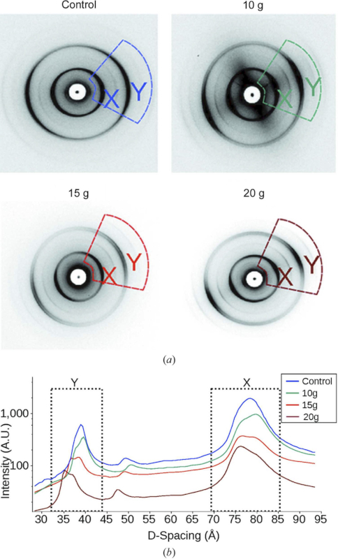
As reported in the Journal of Synchrotron Radiation, investigators from Joseph Orgel at the Illinois Institute of Technology, Argonne National Laboratory and the Army Research Lab studied the effect of controlled amounts of compressive force on rat optic nerves to attempt to identify the changes that occur in otherwise normal looking brain neurons due to the specific impact forces experienced during head trauma. This first-of-its-kind study used x-ray diffraction data obtained at the Biophysics Collaborative Access Team (Bio-CAT) 18ID beamline at the APS to examine the changes to myelin, the fatty material that wraps around nerve cell projections in the brain and other parts of the body. Starting with no force and working upwards, researchers detected the exact force level at which a detectable change in the myelin structure occurred. The changes were subtle, less than a nanometer but they consistently occurred at the same small load of force. What is more, the researchers were able to measure just how much the myelin sheath changed - reflecting the kind of change that occurs in head trauma.
As a result of this ongoing work, researchers have a better understanding of what kind of experience, or injury, leads to what kind of damage in the myelin - helping to visualize injuries based on the smallest force necessary to cause it. This information may be critical to knowing when someone has an injury after an accident but before symptoms emerge, and help supports the decision of when and how to treat them.
See: Orgel J, Madhurapantula RS, Eidsmore A, Wang M, Dutov P, Modrich CD, Antipova O, McDonald J, Satapathy S. “X-ray diffraction reveals blunt-force loading threshold for nanoscopic structural change in ex vivo neuronal tissues,”“. J. Synchrotron Radiation, 2019. 26(Pt 1): p. 89-95. PMCID: PMC6337887, DOI: 10.1107/S1600577518015035.