Relaxation at the Molecular Level

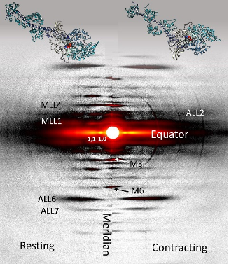
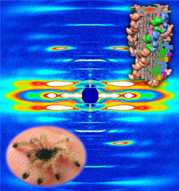
Two-dimensional x-ray diffraction patterns from resting (left) and contracting (right) mouse soleus muscle and the corresponding myosin structures. The differences in myosin structures (scallop myosin) during resting (left) and contracting (right) can be detected by the x-ray patterns. Pattern changes were tracked in live muscle in a time resolved manner.

The molecular interactions between the proteins myosin and actin that generate force during muscle contraction are some of the most well-studied molecular interactions in biology. However, there are some congenital skeletal muscle disorders and types of heart failure where relaxation of the muscle, rather than the force generation part of the cycle, appears to be the problem, and there are currently no available treatments that affect relaxation specifically. A more detailed understanding of the dynamics of the relaxation process could help in the development of treatments that maintain or increase force generation while repairing defects in relaxation. Recent work conducted at BioCAT used a unique transgenic mouse model, time-resolved small-angle x-ray diffraction, and molecular dynamics simulations to discover more about how myosin and actin interact during skeletal muscle relaxation. This research, published in the Journal of Physiology, demonstrates that this type of small-angle x-ray analysis may be of great value …

more ...

Understanding the Physiology of the Human Heart through the Study of Tarantula Muscles

The human heart is a remarkable feat of evolutionary engineering. Beating about 100,000 times per day and pumping nearly 2,000 gallons of blood through an interconnected series of veins, arteries, and capillaries that spans a distance greater than 60,000 miles, the heart is the most important muscle in the human body. Yet, heart disease remains the number one cause of death in the world, demonstrating the need for more research in heart physiology. Now a research team has found an unlikely source of inspiration for understanding how the human heart works and how we might design better drugs for conditions like hypertrophic cardiomyopathy: tarantulas. The source of nightmares for arachnophobes and the household pets for arachnophiles are inspiring researchers to take new approaches to understanding diseases that alter how heart muscle cells contract and relax. But, before getting to the human heart, there is more to learn about the physiology of tarantula muscles. The researchers set out to understand how contractions in tarantula muscle cells are activated and why are muscle twitches that follow a sustained muscle contraction (post-tetanic) more forceful than those that don’t (pre-tetanic). Their results provide evidence that phosphorylation, the chemical addition of …

more ...

Key Insights into an Inherited Muscle Disease

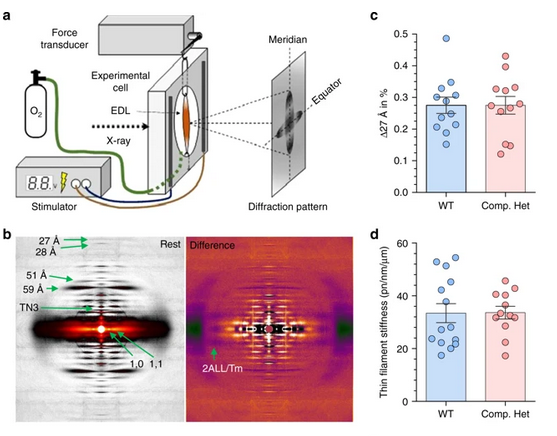
Compound nebulin mutations cause changes in thin filament structure.

The gene NEB encodes for the skeletal muscle protein nebulin. Mutations in NEB cause the disease nemaline myopathy, which is one of the more common inherited myopathies. Patients with this muscle disorder have muscle weakness in multiple different parts of their body and can also experience difficulties with feeding or breathing. Currently, there is no cure for nemaline myopathy and treatment options are limited. Therefore, there is a need to better understand this disease and design new therapeutics that can improve patient quality of life. A team of researchers from the University of Arizona working to provide new insights into the pathogenesis of this skeletal muscle disorder, report a new mouse model of nemaline myopathy that exhibits similar symptoms to those identified in human patients. An important part of this work utilized x-ray diffraction data collected at the APS. The diffraction data provide new insights into how mutations of NEB alter the molecular structure of skeletal muscles. The findings from the study are highly impactful because they significantly increase our understanding of how mutations in the gene NEB cause nemaline myopathy. Importantly, the new mouse model of this disease can be …

more ...

Structure-Function Studies Elucidate GPCR-Independent Regulation of G-proteins

Guanine nucleotide binding proteins popularly known as G-proteins, involved in a variety of cellular signal transduction pathways are heterotrimeric proteins consisting of α, β, and γ subunits. Ric8A is known to be both a chaperone for the assembly of the α-subunit of G-proteins, and a Guanine nucleotide Exchange Factor (GEF). McClelland et al., have conducted a detailed structural analysis on the complex between Ric8A and Gαi1 using cryoEM, X-ray crystallography, and SAXS. Constructs of Ric8A and Gαi1 optimized for structure determination and to reduce conformational heterogeneity were used to assemble the Ric8A-Gαi1 complex. They were able to determine the interface between the two proteins which consists of three separate surface contacts which essentially stabilize Gα in its nucleotide-free state. Furthermore, it was found that a specific Casein Kinase mediated phosphorylation of Ric8A stimulated the GEF activity by structurally stabilizing the Ric8A-Gα interface. Ric8A binding to the disrupted Guanine nucleotide binding site on Gα was determined to be critical for the GEF function as point mutants at residues of Ric8A involved in this interaction compromised the GEF activity. A significant amount of re-organization of a particular helical domain in Gα relative to the GTPase domain is involved in providing a means …

more ...

Uncovering Unique Structural Features in Protein Regions Associated with ALS

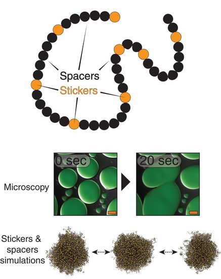
Single chain of the model (top), microscopy of liquid-liquid pase separation (middle), and images from phase separating simulations (bottom).

Many of us are familiar with mad cow disease–the neurodegenerative disease caused by prions. Although they have a similar name, the less familiar prion- like domains (PLDs) refer to something different–unique, low-complexity regions of proteins that are capable of regulating gene expression and affecting important cellular processes. Prion-like domains have become a topic of interest because of their connection with a variety of debilitating brain diseases, such as amyotrophic lateral sclerosis (ALS) and frontotemporal dementia. In fact, mutations in PLDs of some genes have been shown to cause neurodegenerative disease. For example, mutations in PLDs of the genes hnRNPA2B1 and hnRNPA1 can cause the neurodegenerative disorders ALS and multisystem proteinopathy. A recent study using data obtained at BioCAT completed a comprehensive biophysical investigation of PLDs in the protein hnRNPA1 to uncover the major behavioral and structural features of these domains. This meaningful work may lead to discoveries that can help individuals living with such neurodegenerative diseases.

Amyotrophic lateral sclerosis is a devastating disease of the nervous system that affects the brain and spinal cord. Patients often present with symptoms …

more ...

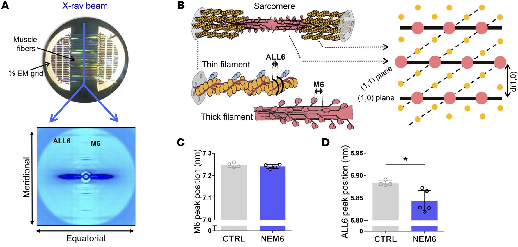
Sarcomere Structure and Nemaline Myopathy

NEM6 mutations cause changes in thin filament structure.

Nemaline myopathy (NM) is one of the most common congenital non-dystrophic myopathies and is characterized by severe hypotonia, muscle weakness, feeding difficulties, respiratory failure, and the presence of nemaline bodies (rods) in skeletal muscle biopsies. One form of nemaline myopathy is caused by mutations in the KBTBD13 (NEM6) gene. In addition to weakness, NEM6 patients have slow muscle relaxation, compromising contractility and daily-life activities. The role of KBTBD13 in muscle is unknown, and the p athomechanism underlying NEM6 is undetermined. A combination of transcranial magnetic stimulation-induced muscle relaxation, muscle fiber- and sarcomere-contractility assays, super-resolution microscopy, and low angle X-ray diffraction on the BioCAT Beamline 18ID revealed that the impaired muscle relaxation kinetics in NEM6 patients are caused by structural changes in the thin filament, a sarcomeric microstructure. Using homology modeling, binding- and contractility assays with recombinant KBTBD13, novel Kbtbd13-knockout and Kbtbd13R408C-knockin mouse models and a transgenic zebrafish model the authors discovered that KBTBD13 binds to actin – a major constituent of the thin filament - and that mutations in KBTBD13 cause structural changes impairing muscle relaxation kinetics. The authors propose that this actin-based impaired relaxation is central to NEM6 pathology.

See: Josine M. de …

more ...

Structure-Function Understanding of aGPCR ECRs Critical for Drug-Design

Cellular communication mediated by a variety of cell-surface receptors involves ligand induced conformational changes in the extracellular region (ECR). A variety of drugs such as cetuximab (Epidermal Growth Factor Receptor), etrolizumab (Integrins), and erenumab (calcitonin receptor-like receptor) function by trapping ECRs in specific conformations and have proved to be effective therapeutic agents in several cancers, bowel diseases, and migraine. Leon et al., have addressed a class of relatively understudied G-protein couple receptors (GPCRs) called adhesion-GPCRs (aGPCRs) which have a structurally unique ECR with a diverse set of mechanistic possibilities. Specifically, they study the Gpr126, which is known to be essential for Schwann cell myelination, involved in heart development in the mouse model, and inner ear development in Zebra fish. Determination of the high-resolution structure of the Zebra fish Gpr126 ECR revealed the involvement of a heretofore undefined domain that has splice variants with or without a 23aa stretch in different isotypes (-ss or +ss) and is the primary determinant of whether the ECR is in the open active state or the closed inactive state. Also remarkable was the calcium dependent site at the tip of the ECR which promoted the closed inactive state of Gpr126. The closed conformation observed in …

more ...

Cool Temperatures During Hibernation May Freeze Muscle Contraction to Save Energy

Low temperatures shift myosin heads from an ordered relaxed state to a disordered state that cannot bind actin in response to stimulation.

Striated muscle contraction is a highly regulated process that involves an orchestrated series of events within the muscle’s contractile units, which are also known as sarcomeres. In a recent study, researchers studied the effect of low temperature on mammalian skeletal muscle contraction. They found that cooler temperatures reduce force generation by trapping filaments in the muscle sarcomeres in a refractory state that cannot undergo contraction and utilize adenosine triphosphate (ATP). This mechanism provides important insight into how hibernating animals may conserve energy while still allowing vital functions in the body to continue.

Muscle tissue’s repeating functional units, the sarcomeres, impart a striated or striped appearance to the tissue when examined under a microscope. Sarcomeres contain thin (containing the protein actin) and thick (containing the protein myosin) filaments that power contraction of skeletal muscle and cardiac muscle, both of which are striated.

Contraction of striated muscle is triggered when calcium enters the muscle fibers. This leads to structural changes in proteins known as troponin and tropomyosin that reside in the thin filament. Ultimately, this allows actin and …

more ...

Frustration and Folding of a TIM Barrel Protein

Multiple folding pathways discovered by simulations, validated against the time resolved SAXS and FRET data.

In their continuing endeavor to understand misfolding proteins as part of the etiology of a variety of diseases, the Matthews lab particularly focuses on the different factors that impede a protein’s path from the unfolded state to the global free energy minimum. The complexity of the folding trajectory understandably depends on the size of the protein mostly because of the formation of intermediates many of which often stall the formation of an optimal native conformation.

The triosphosphate isomerase (TIM) barrel family of proteins have very well characterized and conserved folding path despite large variations in sequence which make it an ideal group of proteins to obtain widely applicable insights into the folding process. Like other proteins studied before, S.solfataricus indole-3-glycerol phosphate synthase (SsIGPS) a TIM barrel protein goes through a burst phase followed by a relaxation phase and then eventually folds into the native conformation. Mutational and hydrogen exchange experiments have helped characterize the species found in the few millisecond time range of the folding process. In this study, Halloran et al., have used a combination of continuous flow FRET, SAXS and simulations …

more ...

Structure of BS Ric8A, a regulator of G-protein Biology

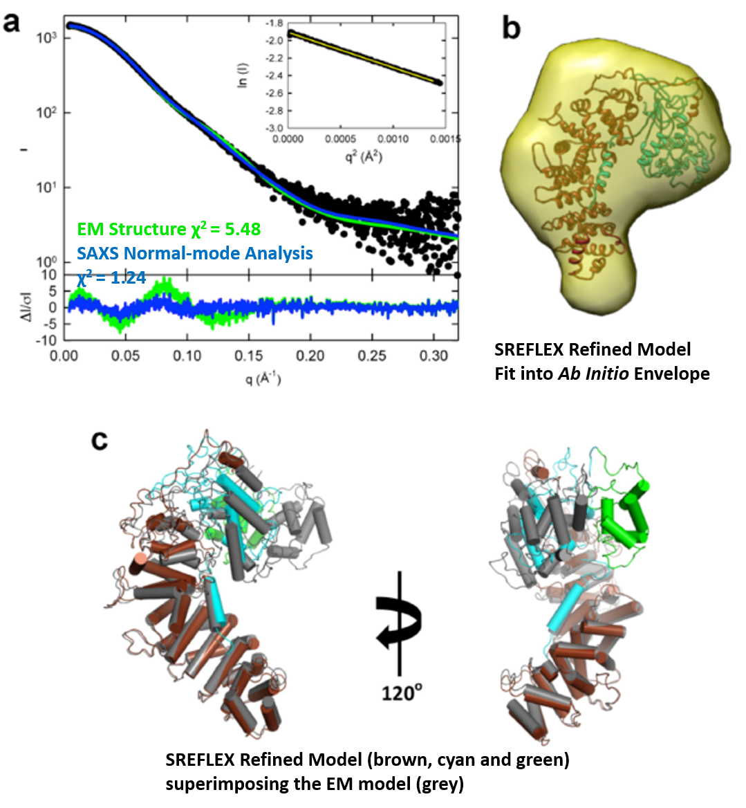
A model of the Ric8a protein structure showing the best scoring flexible tail (right), and the validation of the model against the SAXS data (left).

G-protein signaling has been the dominant theme in the Artemyev lab and their recent work specifically addresses Ric8A (Resistance to inhibitors of cholinesterase 8A) structure and function. Ric8A is a well-known regulator of G-protein biology and belongs to a class of proteins different from the G protein-coupled receptors (GPCRs), which act via interactions with monomeric Gα subunits as opposed to heterotrimeric Gαβγ proteins. SAXS was used in combination with crystallography and biochemical studies to show that the flexible C-terminal tail is important for the overall stability of Ric8A and the function as a guanine nucleotide exchange factor (GEF). The crystal structure revealed that Ric8A belongs to a functionally diverse class of proteins with what is known as an armadillo-fold (ARM) characterized by two layers of alpha helices arranged in a right handed superhelix. Ric8A diverged from the class in terms of the number of ARM repeats (8 as opposed to 10) and is further followed by a flexible region spanning ~ 70 residues.

Differential scanning fluorimetry (DSF) was used to determine that a construct without the …

more ...