An Unusual Shape Change to Deliver Selenocysteine to Proteins

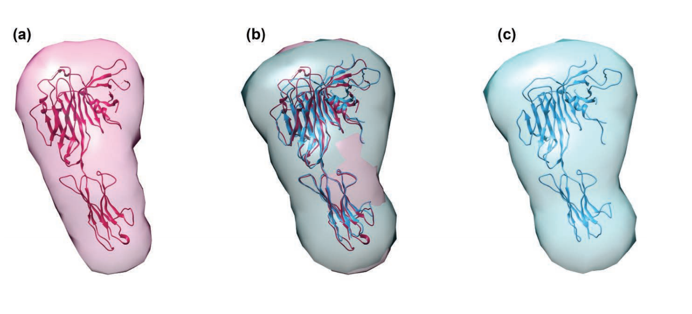
Fig. 1. (A) Cartoon (left) and surface diagram (right) of the overall structure and domain organization of human eEFSec. The color-coding is according to the scheme shown below. (B) The GTP-to-GDP exchange on human eEFSec induces an unexpected conformational change in D4, but not in D1. A comparison of the GTP- (light blue) and GDP-bound states (light red) reveals a lack of the canonical conformational change in the EF-Tu-like domain (D1-3). Instead, D4 swings ~26° towards the dorsal face of the molecule and away from the trNA-binding site. The view is rotated ~90° clockwise relative to that in (A).

The element selenium is incorporated into proteins through the 21st amino acid selenocysteine (Sec). Such selenoproteins are critically important to all types of life, suggesting that being able to accurately decode the Sec codon and correctly placing this amino acid in proteins is biologically fundamental. However, little is known about biosynthesis of selenoproteins in eukaryotic cells. To better understand this process, a team of researchers used data gathered at two APS sectors to determine the crystal structure of the human translational elongation factor responsible for recognizing and delivering the transfer rnA (trnA) carrying Sec to the ribosome …

more ...

TAPBR: A Novel Protein Chaperone With a Role in Peptide Editing in Immune Recognition

Fig. 1. Calculated SAXS envelopes for tapasin (a) and TAPBPr (c), as well as both envelopes superimposed (b). A ribbon diagram of the high-resolution x-ray structure of Tapasin, or of a molecular homology model of TAPBPr, is also superposed on the SAXS envelopes.

TAP binding protein, related (TAPbPr), a novel protein chaperone, plays a role in loading peptides onto major histocompatibility class i (mhc i) molecules during the process of immune surveillance. However, until now, how TAPbPr functions in antigen presentation has been unknown. Researchers investigated the biochemical function of TAPbPr, comparing it with tapasin, another chaperone with a similar protein sequence. Using direct binding studies, they demonstrated that TAPbPr functions as a peptide editor, and binds mhc i molecules that are either peptide-free or complexed with low affinity peptides. Because macromolecular crystallography has so far proven unsuccessful for obtaining high-resolution structural information on TAPbPr, the researchers instead collected small-angle x-ray scattering (SAxS) data at the APS. This allowed them to confirm the structural similarities between TAPbPr and tapasin. The results of this study could lead to ways to modulate peptide loading in vaccine design, improving T-cell recognition.

TAPbPr is involved in the intracellular loading of peptides onto mhc i …

more ...

Lafora Disease: A Delicate Solubility Problem

Our cells are brilliant biochemists that solve all sorts of chemistry problems under difficult conditions. They speed up slow reactions by orders of magnitude, stuff miles of DNA into tiny spaces, and carefully balance the solubility of different kinds of molecules in a jam-packed cellular solution. A good example is the storage of glucose energy in cells in the form of glycogen. Cells can store up to 55,000 glucose units in water-soluble spheres of branched, polymeric glycogen. This provides ready energy for rapid response to cellular needs but also must be managed carefully because too much glycogen accumulation can activate programmed cell death. This is especially true of neurons, which consume large amounts of glucose but are particularly sensitive to glycogen build-up. One example of what can happen when this basic metabolic process goes awry is observed in Lafora disease, a devastating fatal epilepsy in which mutations in a single key enzyme result in the formation of insoluble glucan inclusion bodies that cause neuronal death. Research conducted at two x-ray beamlines at the U.S. Department of Energy’s Advanced Photon Source (APS), an Office of Science user facility at Argonne solved the structure of the enzyme responsible, the …

more ...

Unique Bacterial Chemist in the War on Potatoes

In fertile farm soils where potatoes grow, Streptomyces scabies bacteria wage war using chemicals related to explosives and pesticides.

But a microbial spoiler defuses one of S. scabies’ poisons. Researchers at the Georgia Institute of Technology using high-brightness x-rays from the U.S. Department of Energy’s Advanced Photon Source (APS) have gained new insights into a one-of-a-kind mechanism the microbe employs, which could someday contribute to the development of new agents to degrade tough pollutants and help rescue crops.

When S. scabies infects potatoes, it spews poisons called thaxtomins, which riddle potatoes with familiar dark scabs. Perhaps a trifle to the potato connoisseur excising them with a paring knife, but on a global scale, the blemishes add up to a slash in agricultural production.

Scientists investigating potato soil have found bacteria of the species Bradyrhizobium sp. JS329 running interference. Though their tough enzymes don’t break down thaxtomins, they do render innocuous another S. scabies toxic secretion called 5- nitroanthranilic acid (5-NAA).

Still, understanding how 5-NAA is broken down could prove useful to agriculture. “The 5NAA molecule is similar enough to thaxtomin that studying its degradation might inspire future work to engineer an enzyme or bacterium, or …

more ...

Unearthing the Mechanism of the Frank-Starling Law, A Central Regulator of Heart Function

Fig. 1. A typical two-dimensional x-ray diffraction pattern of heart muscle, obtained at the Biophysics Collaborative Access Team (BioCAT) beamline at the APS. meridional reflections (horizontal, yellow) arise from thin and thick filament proteins. Stretching of these fibers can change the spacing and/or intensity of these reflection, allowing researchers to observe changes within the heat’s muscle on a nanometer scale.

The Frank-Starling law of the heart is a basic physiological principle first observed more than 100 years ago. it describes how the heart is able to move blood through the body in a regulated way by pumping out as much blood as it receives. To understand the nature of the molecular mechanism underlying this important regulatory process, researchers from Loyola University together with colleagues from the Illinois Institute of Technology and the University of Wisconsin–Madison conducted x-ray diffraction experiments at the APS to examine myocardial muscles of rats deficient in length-dependent activation (ldA) of muscle fibers in the heart. Their work shows that the protein titin is critically important for transmitting the stretch-induced signals within the heart’s muscles known to impact the strength with which the heart contracts. This work not only solves a piece of …

more ...

Identifying the Structure of a Tumor-Suppressing Protein

An activated PTEN dimer that contains two non-mutant proteins (A) can transform the functional lipid (D) on the cellular membrane (E) into a chemical form that tunes down cancer predilection. Dimers that contain a mutated protein (B), or PTEN monomers cannot transform the functional lipid. Image: Carnegie Mellon University

The dimer structure of an important tumor-suppressing protein, phosphatase and tensin homolog (PTEN), the second most frequently mutated protein found in human cancer, has been established by an international group of researchers carrying out studies using the BioCAT beamline 18ID at the U.S. Department of Energy’s Advanced Photon Source (APS), an Office of Science user facility. Their findings provide new insights into how the protein regulates cell growth and how mutations in the gene that encodes the protein can lead to cancer. The study was published online in Structure, and appeared in the October 6, 2015 issue.

Phosphatase and tensin homolog is a known tumor suppressing protein that is encoded by the PTEN gene. When expressed normally, the protein acts as an enzyme at the cell membrane, instigating a complex biochemical reaction that regulates the cell cycle and prevents cells from growing or dividing in an unregulated fashion. Each …

more ...

Unraveling Protein Folding

For many proteins, the ability to change shape is essential for their proper functioning within cells. One longstanding question concerns the process proteins follow when shifting from an unfolded to a three-dimensional globular form. Most previous studies have supported the idea that when an unfolded protein is exposed to native conditions (i.e., to an environment favoring its typically fully-folded, physiological form) a continuous unfolded-to-semi-folded collapse ensues. However, other studies suggest that there is an energetic bottleneck to this step that renders it an all-or-none transition. To resolve the issue, researchers from the University of Massachusetts Medical School and the Illinois Institute of Technology probed the folding stages of cytochrome c, an archetype for protein folding behavior. Its microsecond-scale folding dynamics were unambiguously characterized with Förster resonance energy transfer (FRET) complimented by small angle x-ray scattering (SAXS) carried out at the APS. The SAXS and FRET measurements refuted the conventional view that cytochrome c folding proceeds immediately and smoothly when exposed to native conditions; instead, subpopulations of unfolded and semi-folded protein were observed to coexist during brief intervals. These results provide fundamental insights for biochemistry, as well as for human diseases characterized by protein misfolding, including Parkinson’s, Huntington’s …

more ...

A Molecular Basis for Protease’s Role in Preventing Alzheimer’s Disease

Fig. 1. (a) hPreP is comprised of an N-terminal domain (purple) and C-terminal domain (blue), which enclose the catalytic chamber and active-site (green) (b) hPreP captures Ab using an exosite (dashed box; D) and active site (dashed box; C) (c) Close-up of the hPreP active site (blue mesh), bound to Ab (gray) prior to cleavage between Ab residues F20 and A21. The Ab peptide is trapped in the catalytic site by the S1 site (indicated) (d) Closeup view of Ab (blue mesh) anchored in the hydrophobic hPreP exosite (shaded gray). Adapted from J.V. King et al., Structure 22, 996 (July 8, 2014).

Proteases are specialized enzymes responsible for degrading damaged, misfolded, or unneeded proteins within the cell. The human mitochondrial presequence protease (hPreP) breaks down several distinct proteins, including beta amyloid (Aβ) species known to aggregate and form the amyloid plaques associated with Alzheimer’s disease. Using a combination of high-resolution x-ray crystallography and small angle x-ray scattering (SAXS) at the APS, researchers from The University of Chicago and the University of Illinois at Chicago were able to define the mechanism by which hPreP recognizes a diverse array of amyloid proteins. The findings reveal that hPreP uses a large …

more ...

Combating an Infectious Invader

Fig. 1. X-ray fiber diffraction patterns of fungal prion HET-s(218-289). The pattern on the left represents the wild-type with two-rung β-solenoid diffraction while the pattern on the right represents a mutant version of HET-s with stacked β-sheets that are not in the β-solenoid structure.

How does one kill an infectious invader that is not technically alive? That question has vexed researchers studying prions—aggregates of aberrantly folded protein that propagate by inducing properly folded proteins to convert into misfolded, disease-associated forms—since these infectious agents were identified as the source of a range of devastating neurological diseases such as transmissible spongiform encephalopathies (TSEs), which include Creutzfeldt-Jakob disease and “mad cow disease.” Prions contain no genetic material, so their structure determines their biological activity. That structure, made rigid and insoluble through aberrant protein folding, also makes prions difficult to combat. Researchers used two U.S. Department of Energy synchrotron light sources including the APS to pry out new structural information about prions, yielding insights into the fundamental mechanisms of prion at a level of complexity that has not been observed in short-peptide amyloids used as prion model systems.

Unlike viruses, bacteria, fungi, and parasites, prions do not spread …

more ...

X-ray Florescence Imaging: A New Tool for Studying Manganese Neurotoxicity

Mn and Fe distributions in the choroid plexus. XRF images of the Fe (A) and Mn (B) distributions in the choroid plexus (CP) within the lateral ventricle (lv) as identified by increased Fe signal. Images are Bregma −0.48 mm coronal sections of untreated rats (top) and Mn treated (bottom). Yellow dashed lines indicate the boundary between the ventricle and the labeled structures (CPu and HPC, hippocampal formation). The Fe signal shows the presence of CP (containing blood) within the ventricle. Mn concentration in the ventricle is lower than in adjacent brain structures of the CPu and HPC indicating clearance of Mn from the CP. All values given are in µg/g. Scale bar represents a length of 2 mm.

Manganese (Mn) is an essential element required in trace amounts for proper body function. However, despite its vital role in enzymatic reactions, excessive Mn exposure leads to a condition known as manganism or Mn induced parkinsonism. Clinical signs and symptoms of manganism closely resemble those of Parkinson’s disease (PD) and both diseases are pathologically associated with damage to the basal ganglia. While this condition was first diagnosed about 170 years ago, the mechanism of the neurotoxic action of Mn …

more ...