How Water Molecules are Connected

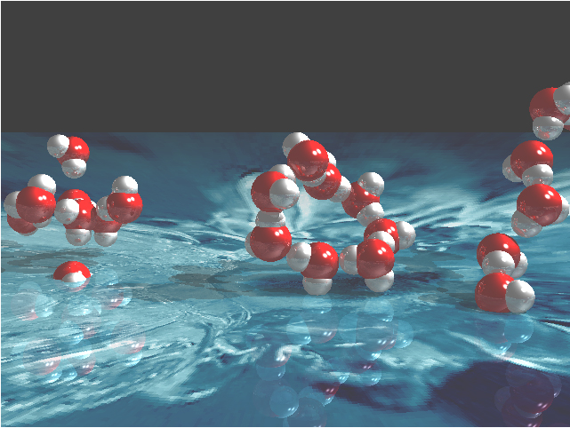
Water may be the most important molecule on Earth, but our understanding of its properties is embarrassingly limited. In solid ice form, water takes on numerous phases and structures that can be studied by means of diffraction techniques. As a liquid, however, water poses a frustrating structural puzzle because of the complex hydrogen bonding that forms a disordered network. Recently, researchers from the Stanford Synchrotron Radiation Laboratory, the BESSY laboratory, Stockholm University, Linköping University, and Utrecht University have used the BioCAT 18-ID beamline at the APS, as well an Advanced Light Source (ALS) beamline, to obtain detailed information about the nearest neighbor coordination geometry in liquid water.

Previous experimental efforts to understand water structure have relied on several methods including infrared spectroscopy and neutron and x-ray diffraction. Unfortunately, the structural information provided by infrared spectra is ambiguous for water, and diffraction provides only radial distribution functions that do not allow unique assignment of local hydrogen bonding configurations. In the work reported by Wernet et al., x-ray absorption spectroscopy (XAS) and x-ray Raman spectroscopy (XRS) were used to investigate local bonding in the first coordination shell of water. In XAS, x-rays are absorbed by core electrons close to the nucleus …

more ...

Grasping the Structure of Insect Muscle Poised to Contract

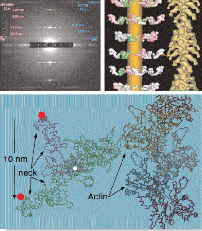
Fig. 1. Top left: Low-angle x-ray diffraction pattern from relaxed insect flight muscle. (Pink and blue numbers represent spacings between repeating structural elements as calculated from the pattern.) Top right: Structure (left) of the modeled myosin heads (pink/white/green) in their relaxed, pre-powerstroke state and (right) in their “rigor” or post-powerstroke state. Bottom: Transition from the head shape in relaxed insect muscle (blue) to rigor-like final shape (green), showing the pivots needed (red on thick filament, white within head) to let the neck swing from one form to the other when bound to actin.

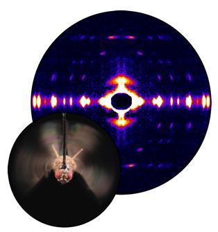
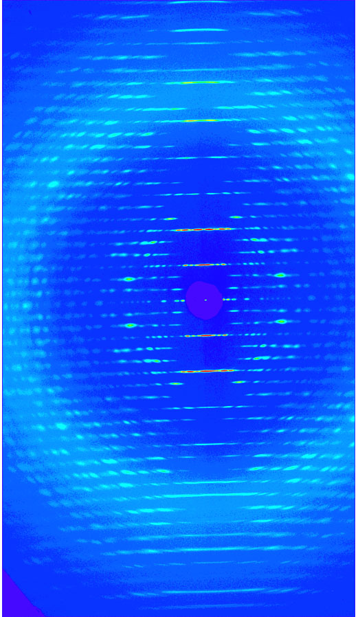
Researchers have achieved the first detailed view of resting muscle filaments poised to contract, a long sought window into the biochemical cycle that causes muscle contraction. The group determined the overall structure of insect muscle fibers from x-ray diffraction patterns and performed computer modeling to analyze the data. The resulting structure, reported in August 2003, suggests a specific mechanism for insect muscle activation.

Muscle cells (fibers) contain two parallel, overlapping sets of protein filaments, made of myosin and actin, respectively, that align with the direction of contraction. The myosin and actin filaments have fixed length individually, but they slide past each other like telescope segments …

more ...

Role of Interfilament Spacing in Muscle Filament Calcium Sensitivity

The heart regulates ventricular output in response to changes in ventricular filling, a mechanism known as Frank Starling’s law of the heart. Part of the cellular basis for the law is an increase in myofilament Ca2+ responsiveness upon an increase in sarcomere length (SL). (Sarcomeres are the basic functional units of muscle fibers, which contract in response to calcium influx.) How information on SL is transmitted to myofilaments is unclear. One prominent hypothesis is that a decrease in myofilament lattice spacing (LS) in response to an increase in SL underlies the Ca2+ sensitivity.

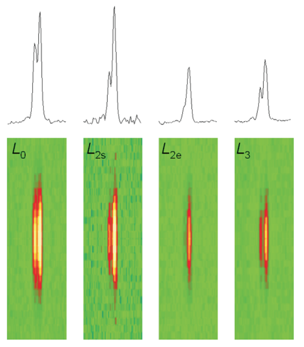
In support of this hypothesis, shrinkage of the myofilament lattice by osmotic compression using high-molecular-weight moieties has been shown to increase myofilament Ca2+ sensitivity at a short SL, mimicking the effect of increased SL. The extent of LS shrinkage in that experiment was estimated from the reduction in muscle width, however. Recently, researchers directly measured LS by synchrotron x-ray diffraction as a function of SL in skinned rat cardiac trabeculae bathed in 0-6% dextran (high-molecular-weight) solutions.

To assess the effect of SL on myofilament Ca2+ sensitivity, Ca2+-contraction force relationships were determined at three SLs (1.95, 2.10, and 2 …

more ...

The “Second Stalk” of ATP Synthase: Dimerization Domain Structure

Fig. 1. Function and structure of the b2 dimerization domain in ATP synthase. ATP synthesis is catalyzed as follows. Proton flow through the membrane-embedded F0 portion causes the ring of c subunits and the γε “central stalk” to rotate, which bridges the F0 and F1 complexes. As the central stalk rotates, it drives conformational changes in the β-subunits. The “second stalk” of the complex is composed of the b2 and δ subunits. Its function is to provide a stator to prevent rotation of F1. A model for the dimerization domain of b2, derived from the crystal structure of the b62-122 monomer and SAXS of the solution dimer, is shown to the right of the ATP synthase holoenzyme. The alanines that define the undecad repeat, characteristic of a right-handed coiled coil, are in green.

Hydrolysis of adenosine triphosphate (ATP) drives many of the vast range of energy-consuming processes within a cell. The ATP synthase enzyme is a molecular motor that couples proton movement to ATP synthesis. In the rotational model of the enzyme, protons flow down an electrochemical gradient, through the membrane-embedded F0 portion of ATP synthase, causing a ring of c …

more ...