Homomeric interactions of the MPZ Ig domain and their relation to Charcot-Marie-Tooth disease

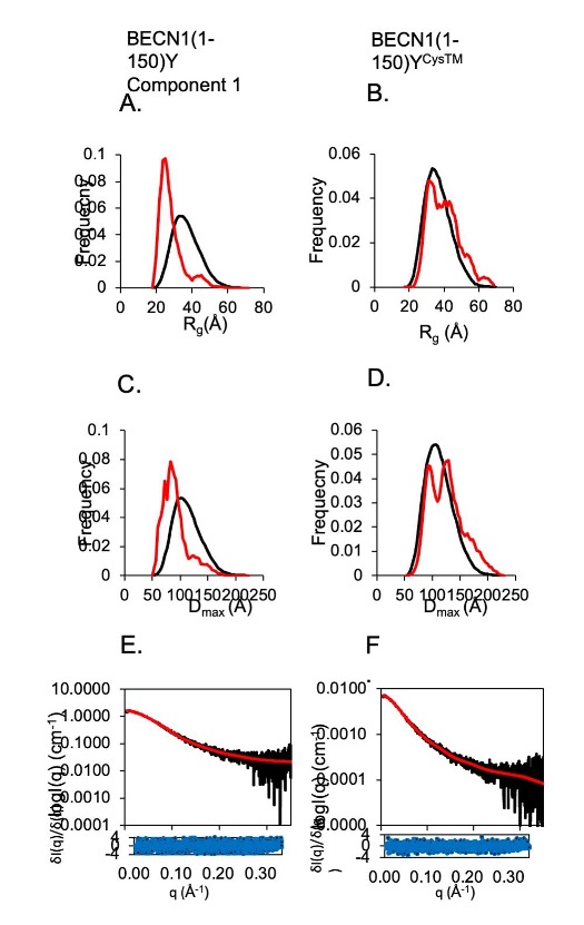
Tetrameric building blocks are integral to the current models for IgMPZ functional assembly in Myelin. The authors used SAXS to identify and characterize the tetramerization interface.

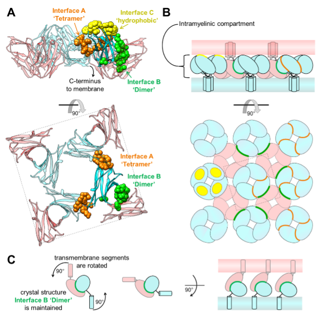
Charcot Marie Tooth (CMT) disease is the most common form of heritable peripheral neuropathy, which are a group of inherited diseases affecting the peripheral nervous system (PNS). Myelin protein zero (MPZ) is necessary for normal myelin structural and function comprises ~50% of all proteins in the PNS; mutations in MPZ account for around 5% of CMT cases. MPZ is a transmembrane adhesion protein which holds together adjacent myelin membranes, thought to be mediated in part through homotypic interactions of its extracellular Ig domain. Exactly how the Ig domain of MPZ (IgMPZ) mediates adhesion of apposing membranes is not yet fully understood but is nonetheless important for understanding how myelin is constructed and how mutations in the IgMPZ cause disease. Models for how the IgMPZ might form oligomeric assemblies has been extrapolated from a protein crystal structure in which individual rat IgMPZ subunits packed together to form 3 weak interfaces involving IgMPZ organized tetramers, a ‘dimer’ interface linking tetramers together, and a hydrophobic interface that mediates binding to lipid …

more ...

Right Ventricular Sarcomere Contractile Depression and the Role of Thick Filament Activation in Human Heart Failure With Pulmonary Hypertension

Right ventricular (RV) contractile dysfunction commonly occurs and worsens outcomes in patients with heart failure with reduced ejection fraction and pulmonary hypertension (HFrEF-PH). However, such dysfunction often goes undetected by standard clinical RV indices, raising concerns that they may not reflect aspects of underlying myocyte dysfunction. To address the need for better diagnostics, the authors sought to characterize RV myocyte contractile depression in HFrEF-PH, identify those components reflected by clinical RV indices, and uncover underlying biophysical mechanisms.

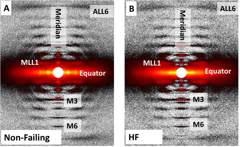
X-ray diffraction and myosin ATP turnover quantification assays show that patients with HFrEF-PH RV dysfunction with depressed isometric tension have reduced on state and disordered-relaxed (DRX) myosin; moreover, increasing the proportion of DRX myosin rescues the RV failure myocyte phenotype, whereas depressing DRX induces it. These results show, for the first time, that reduced basal DRX (on state) myosin contributes to a heart failure phenotype. HFrEF-PH RV myocytes exhibit blunted stretch-mediated recruitment of myosin from its super-relaxed to its DRX state, in association with depressed length-dependent active stiffness, a key contributor to Frank-Starling reserve. Although there are many RV myocyte contractile deficits in HFrEF-PH, commonly used clinical indices only detect reduced isometric calcium-stimulated force, which is related to …

more ...

Proteins in Heart Muscle Can Produce More Oomph than Previously Thought

more ...

GRB2 dimerization mediated by SH2 domain-swapping is critical for T cell signaling and cytokine production

The SH2/SH2 domain-swapped GRB2 dimer matched the experimental SAXS data significantly better than other models, providing direct structural evidence for this conformation in solution.

Adaptor proteins are accessories to main proteins in signal transduction pathways that usually lack intrinsic enzymatic activity but instead facilitate the linking of binding partners together to enable the formation of larger signaling complexes. One widely expressed adaptor protein is the growth factor receptor-bound protein 2 (GRB2), which facilitates formation of cytoplasmic signaling complexes from a wide array of binding partners, including (among others) growth factor receptors, cytokine receptors and T cell receptor (TCR) ζ chains. As a consequence, the structure and function of GRB2 have become major areas of investigation for novel areas of interventions against various human diseases. GRB2 has been shown to exist in either a monomeric or dimeric state, where GRB2 dimers are formed by the exchange of protein segments between domains, a process termed “domain-swapping”. Prior to this work, swapping between the SH2 and SH3 domains of GRB2 has been demonstrated in the full-length structure, but SH2/SH2 domain-swapping had not. Researchers at the University of Iowa generated a model of full-length GRB2 dimer with an SH2/SH2 domain-swapped conformation …

more ...

Titin force in muscle cells alters lattice order, thick and thin filament protein connections

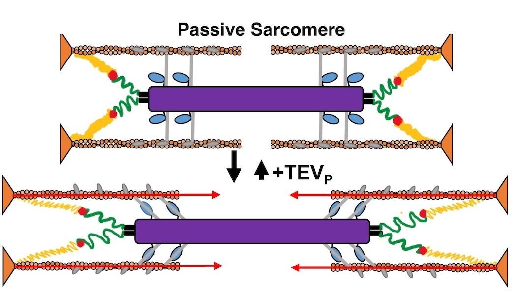
Muscles can produce more force when stretched to a longer length at the same level of activating calcium, a poorly understood phenomenon known as myofilament length dependent activation (LDA). It was suggested several years ago that passive force generated by the giant elastic protein titin could be the length sensor behind this phenomenon, but direct evidence has been lacking. Investigators from the University of Muenster in Germany, Northern Arizona University, University of British Columbia, and the Illinois Institute of Technology used a mouse model with a cleavage site inserted into the titin protein allowing titin to be enzymatically cut in the mature tissue (with TEVp protease) to make it ineffective. Small-angle X-ray diffraction of muscle from these mice with and without cleavage showed that titin cleavage diminished the changes in length-dependent structural signatures (“priming”) associated with LDA. Strikingly, a titin-sensitive, length-dependent structural changes were also seen in thin filaments, which seems only possible if there are bridging structures between the thick and thin filaments in resting muscle, potentially comprised of myosin-binding protein C. These experiments firmly established titin as the length sensor in LDA and showed that LDA involves structural changes in both thick and thin filaments.

See: Anthony L …

more ...

Understanding Phase Separation Could Impact Treatment of Neurodegenerative Disease

Cartoon highlighting the physicochemical contributions of different forces driving phase separation in prion-like low-complexity domains (PLCDs). From A. Bremer et al., Nat. Chem. 14, 196 (February 2022). © 2022 Springer Nature Limited

Living cells are amazing little biochemical factories that conduct countless chemical reactions in a cellular soup packed with lipids, proteins, nucleic acids, and ions, keeping them all in their proper places at any given time. Cells maintain this organization even while carrying out complex tasks such as cell division, signaling, transcriptional regulation, and stress responses. One example of this is the careful management of stress granule formation, a process in which membraneless organelles transiently form to control the utilization of mRNA during stress. These granules form and disperse through reversible liquid-liquid phase transitions involving proteins and RNA in the granules. Recent research has demonstrated that RNA-binding proteins in these granules contain intrinsically disordered sequences, called prion-like low-complexity domains (PLCDs), that are critical to regulation of these reversible phase transitions. There is also mounting evidence that these transitions may be disrupted in neurodegenerative diseases, like amyotrophic lateral sclerosis (ALS), in which mutations in PLCD-containing proteins, such as hnRNPA1, have been implicated as a cause of the disease. Recent …

more ...

Targeting Cancer at the Level of DNA Expression

Small-angle x-ray scattering-refined molecular models of oncogenic promoter G4s. Shown are best-fitting atomic models of the various higher order G4 promoter sequences from c-Myc, k-Ras, and c-Kit promoters displayed with transparent molecular surface representations.

The last 20 years have brought a revolution in targeted therapies for cancer. Small-molecule inhibitors and monoclonal antibodies that target a specific aberrant protein in tumors have provided cancer patients with treatments that are associated with fewer side effects and longer survival than conventional chemotherapy. This has been, in large part, the result of intensive research into the role of oncogenes in cancer development. Oncogenes are normal cellular genes that have become mutated in such a way that they aberrantly promote the uncontrolled cell growth seen in cancer. They are often proteins involved in growth control or activation of cellular signaling; inhibiting these mutated proteins has proven to be effective in stopping the growth of many cancers. Research by a team from the Brown Cancer Center at the University of Louisville in Kentucky using the U.S. Department of Energy’s Advanced Photon Source (APS) and published in the journal Nucleic Acids Research promises to extend these treatment possibilities to control these oncogenes at the gene …

more ...

Understanding the Structural Implications of Genetic Mutations in Heart-Muscle Disease

Mechanism of action for HCM-D166V and DCM-D94A mutations. The HCM-D166V model disrupts the SRX state and promotes the SRX-to-DRX transition increasing the number of DRX heads and leading to hypercontractile behavior. The DCM-D94A model stabilizes the SRX state yielding fewer heads available for contraction and leading to clinical hypocontractility. Abbreviations: ELC, myosin essential light chain; RLC, regulatory light chain; DRX, disordered relaxed; SRX, super-relaxed.

Cardiomyopathies are diseases of the heart muscle in which the muscle of the pumping chamber (ventricle) can become enlarged (dilated cardiomyopathy; DCM) or thickened (hypertrophic cardiomyopathy; HCM), potentially leading to heart failure. There are currently no effective treatments but the disease often has a genetic component related to mutations in the heart muscle proteins that are involved in muscle contraction, so some researchers have focused their therapeutic development efforts on correcting these muscle contraction problems based on the structural basis of the defect. A recent study from a team of researchers using the U.S. Department of Energy’s Advanced Photon Source (APS) employed humanized mouse models expressing mutations observed in patients with HCM and DCM to evaluate the structure-function relationships and the changes observed in cardiac muscle contraction with …

more ...

New Resource for the Muscle Diffraction Community

Three possible conformations of an intrinsically disordered protein: collapsed (purple), expanded (gold) and a combination of collapsed and expanded (red). Image created by Kristina Davis, University of Notre Dame.
BioCAT staff have just published a review article, Ma & Irving, 2022 Int. J. Mol. Sci. 2022, 23(6), 3052, on the use of small angle X-ray fiber diffraction for studying skeletal and cardiac muscle disease. The article consists of a guided tour of the various diffraction features that can be used to extract specific pieces of information that can be used to provide insights into the structural basis of pathology. The article also contains a comprehensive review of the literature reporting diffraction studies of muscle that illustrates how small angle fiber diffraction has increased our understanding of specific muscle diseases such as hypertrophic cardiomyopathy, dilated cardiomyopathy, and nemaline myopathy.
more ...

What Bacterial Pathogens Can Teach Us about Protein Folding

Three possible conformations of an intrinsically disordered protein: collapsed (purple), expanded (gold) and a combination of collapsed and expanded (red). Image created by Kristina Davis, University of Notre Dame.

Protein folding is one of the fascinating unanswered questions in biology. How does an amino acid sequence that is unfolded when it leaves the ribosome manage to fold properly into a highly ordered, lightning-fast enzyme or sturdy structural protein? Why don’t all the proteins in the cell instead just stick to each other, aggregating into a big mess? A unique model system in bacteria may hold some of the answers to these questions. The system involves the study of what are termed autotransporter proteins, which pathogenic bacteria secrete as virulence factors for infection. These proteins are synthesized in the bacterial cytoplasm and cross one membrane into the bacterial periplasm. Autotransporter proteins then remain in an unfolded state in the periplasm until they pass through the outer bacterial membrane, folding properly along the way. This highly specialized protein folding process has attracted the attention of a team of researchers who have used this bacterial system as a model to determine what allows these unique proteins to maintain their disordered state in …

more ...