A “Copper Bullet” to Kill Cancer

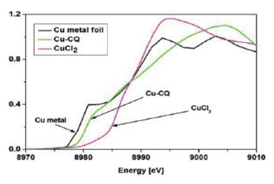
Fig. 2. . The Alzheimer’s drug clioquinol kills cancer cells in the presence of copper by inactivating a key cellular enzyme complex. Shown here are differences in x-ray absorption between copper foil (black line), copper chloride (purple), and copper complexed with clioquinol (green).

A drug normally used to treat Alzheimer’s disease may act as a “copper bullet,” killing tumor cells by coating itself in copper ions, according to research derived in part from studies at the APS. Researchers from Wayne State University, the Henry Ford Hospital, the Illinois Institute of Technology, and Shandong University using BioCAT beamline 18-ID-D at the APS found that the drug clioquinol, when mixed with copper (Fig. 1), killed two types of prostate cancer cell in Petri dishes. The drug without copper also slowed the growth of prostate tumors implanted in mice by up to two-thirds, apparently by soaking up copper ions (charged atoms) present in the implanted tumor cells.

Prostate cancer struck approximately 219,000 U.S. men in 2007 and killed some 27,000 of them, according to National Cancer Institute estimates.

Many types of cancer typically show high levels of copper, including tumors of the prostate, breast, colon, lung, and …

more ...

Filling the Gaps in Collagen Structure

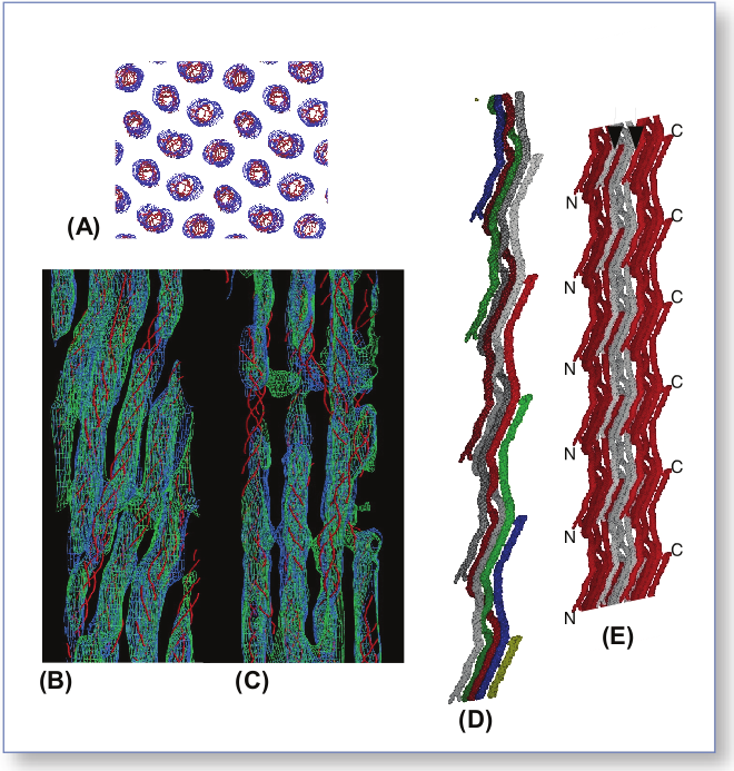
Fig. 1. . . (A) Cross-section of multiple unit cells of the crystalline lattice. (B) Overlap region of the 1-D repeat, with the triple helical backbone of the pre-refined model (red). (C) Same as B, except for the gap region where the super-twist occurs. (D) Two microfibrils, colored differently to easily identify the helical nature of the microfibril twist. (E) Same as D, except three microfibrillar structures are shown, packed together to reveal the interdigitation.
Fig. 2 .. . Background subtracted off-meridian diffraction pattern of rat-tail tendon (left) and simulated diffraction pattern from model-derived intensities (right); the similarity between patterns shows the accuracy of the final model.

Collagens—we might take them for granted, but without them there would be no way to build tissues of the heart, skin, cornea, or bones. In much the same way that wood is used to frame a house and form a structure for the overlying construction materials, collagens are proteins used in the framing of mammalian tissues, but gaining an accurate picture of their three-dimensional structure in the body has proven more difficult. Knowing more about the structure of collagens could help biochemists improve their understanding of heart disease and cancer. Thanks to work by a research …

more ...

Sarcomere Synchrony

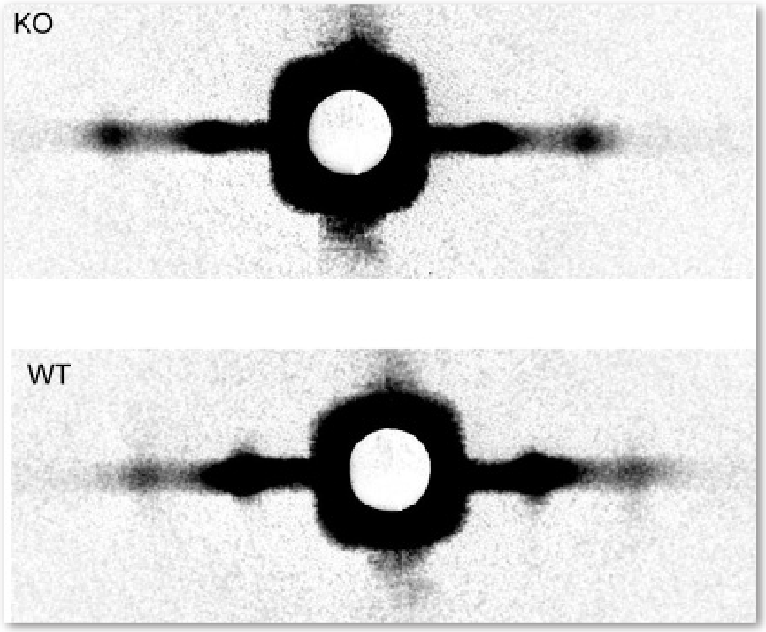
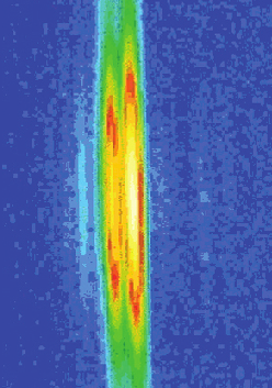
Fig. 1. . Reflection (shown in false color) from an intact contracting frog thigh muscle.

Any time we move our arms, billions of protein molecules work in unison to contract one muscle while letting another muscle relax. The mechanical details of muscle contraction have been known for a long time, deduced from experiments with frog legs, reconstruction of the proteins involved in crystallographic studies, and many other kinds of research. But no one has ever taken such detailed time-lapse molecular pictures of the proteins at work within living muscle. Researchers from Brandeis University, the University of Florence, and Illinois Institute of Technology investigated whether the muscle-contracting proteins work the same way inside muscles as they do inside laboratories. But muscle cells are full of water and the proteins don’t line up quite as regularly as they would purified in crystals. So rather than spend 30 to 40 hours collecting data with a typical x-ray tube, the researchers turned to the BioCAT beamline 18-ID at the APS. With a million times more intensity than a “home source” x-ray tube—and the ability to focus the x-ray beam into a narrow line—the team got a detailed view of the proteins …

more ...

The Correct Signals to Regulate Assembly in Bacteria

You are what you eat” is just another way of saying that input determines output, after some metabolic messing around in between. It’s the “in between” that interests biologists, because that is where the difference between healthy and diseased cells can originate. If the normal sequence of intermediate steps between the beginning and end of a biochemical pathway is disrupted, the disruption can lead to major changes in cellular functioning. A better understanding of the input-output relationships is critical to medical progress. The steps necessary for producing the right output can be numerous and complex, and some pathways have taken decades to unravel. In one of the identified mechanisms, called “two-component signal transduction,” the input causes a receiver domain to be phosphorylated, a step that governs what happens to the output modules. By employing x-ray scattering and electron microscopy researchers from Pennsylvania State University; the University of California, Berkeley; Lawrence Berkeley National Laboratory; The University of Georgia; and the Illinois Institute of Technology using the BioCAT 18-ID beamline at the APS were able to describe—in stunning detail—a novel two-component mechanism for assembling a protein associated with bacterial transcription. Their work greatly advances our understanding of what …

more ...

Reaching for Answers to Questions about the Heart

Michael Reedy (Duke University) and Tanya Bekyarova (Illinois Institute of Technology) are shown in action on the BioCAT beam line 18-ID at the U.S. Department of Energy’s Advanced Photon Source (APS), Argonne National Laboratory. They are studying mechanisms of stretch activation in insect flight muscle.

Stretch activation is very important in human heart muscle when contraction of one part of the heart stretches adjacent muscle tissue causing it to respond a moment later by generating more force. This action aids cardiac “ejection,” i.e., the amount of blood pumped per beat. Muscle structure and function can be studied using high-brilliance x-ray beams, such as those produced by the APS, and the experimental technique time-resolved small-angle fiber diffraction.

Unfortunately, cardiac muscle is relatively poorly ordered and the diffraction patterns relatively uninformative. Fortunately, because of its physiological properties, the flight muscle of insects can be a good model system for cardiac muscle. And because of its high degree of structural order, insect flight muscle produces rich and informative (not to say beautiful) diffraction patterns (image lower right). Muscle contracts by the cyclical action of motor proteins called “myosin” that bind to particular sites on another protein called “actin.” This …

more ...

What Connects Rat Tails to Cancer and Heart Disease?

Collagen is the main (and most abundant) protein in all mammalian connective tissues, including those of the heart, lungs, skin, and tendons. It is also the primary protein in bones and teeth. Bodily malfunctions involving this protein can lead to heart disease and cancer. We know a good deal about how collagen is produced in the body, how it regenerates, and about its physiological structure. But because the collagen protein is so large and insoluble, solving its three-dimensional molecular structure has long been regarded as an impossible, but important, problem. Now, innovative synchrotron x-ray research techniques have yielded new information on the molecular structure of collagen. Because this ubiquitous protein is involved in cancer and heart disease, the data obtained in this study may help in the fight against these deadly ailments.

By adapting methods commonly used for studying much smaller and simpler proteins, researchers from the Illinois Institute of Technology, the Rosalind Franklin University of Medicine and Science, the University of Stirling, and Cardiff University, using the BioCAT, SBC-CAT, and SER-CAT beamlines at the APS, have successfully determined the molecular structure of collagen while it is still intact and undisturbed within whole tendons removed from rat tails …

more ...

Storing the Power to Fly

Fruit flies beat their wings faster than their cellular powerplants can generate the energy needed for flapping. To resolve this energetic discrepancy, researchers from the California Institute of Technology, the Illinois Institute of Technology, and the University of Vermont used the BioCAT beamline 18-ID at the APS to obtain a series of x-ray photographs that revealed the flies’ secret: A muscle protein used to power wings acts like a spring, storing energy while stretched before snapping back. Not only did this finding surprise researchers who study muscle, but the results might also help scientists better understand the human heart.

Drosophila, the fruit fly, beats its wings up and down once every 5 milliseconds. Two layers of muscle control this action: when one layer contracts, it stretches the other. The stretched muscle is then primed to contract; when it does, it stretches the first layer and completes the cycle. These cycles occur faster than nerve impulses can stimulate them; hence, the muscles themselves must keep the beat going.

To find out how muscles do this, the research team needed to visualize the proteins that make muscle contract. Within the flight muscle cells, two proteins cooperate to do this. Myosin proteins …

more ...

Atomic Models of Plant Viruses

Amy Kendall (left), Michele McDonald, and Sarah Tiggelaar — all with the Stubbs Lab in the Department of Biological Sciences at Vanderbilt University — taking data at the BioCAT 18-ID beamline.

Kendall: “Our lab uses fiber diffraction data from oriented sols and dried fibers to determine the structures of flexible filamentous plant viruses. Our long-term goal is to produce atomic models based on diffraction data at resolutions close to 3 Å, but a great deal of information can be obtained at lower resolution, including the size, shape, and symmetry of the viruses.

These viruses are of great significance as models for fundamental virology and cell biology, They have enormous potential as vectors in biotechnology and they are of considerable importance because of the damage they cause to agriculture. We also use the filamentous plant viruses as important model systems for developments in fiber diffraction.”

more ...

Measuring the Efficiency of the Myosin Motor at High Load

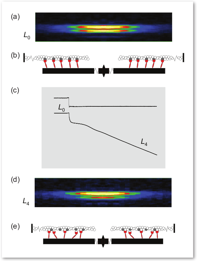
Fig. 1. Two-dimensional (2-D) patterns in the region of the M3 reflection recorded on a charge-coupled device detector at the Bio-CAT beamline with 3-m camera-to-sample distance, normalized by X-ray exposure time. Total exposure times: L0, 25 ms; L2s, 5.5 ms; L2e, 21 ms; and L3, 25 ms. Fiber cross-sectional area equals 38•103 μm2; isometric tension equals 230 kPa; sarcomere length equals 2.16 μm. On the top are shown the intensity profiles obtained by integrating 80 pixels on each side of the meridional axis of the 2-D patterns.

The sliding filament model of muscle contraction is more than 50 years old, yet theories about the precise mechanisms of the motor function still generate some controversy. A group of researchers from Università di Firenze, Istituto Nazionale di Fisica della Materia, Brandeis University, King’s College London, the European Synchrotron Radiation Facility, Illinois Institute of Technology, and Argonne National Laboratory used insertion device beamlines at BioCAT at the APS and at the European Synchrotron Radiation Facility (ESRF) to take a closer look at the molecular structure of myosin II, the molecular motor in muscle, as it works under different loads. This study provides …

more ...

Finding Active Proteins

When combinatorial chemistry produces new varieties of reagents, the tricky next step is figuring out whether those molecules will be biochemically active. While there are several methods for finding active molecules, they all have limitations. Researchers from Argonne National Laboratory, using the BioCAT beamline 18-ID, employed wide-angle x-ray scattering (WAXS) to develop a method for identifying drug candidates.

One way to identify active molecules is to design an assay for the desired function of a protein target and then test each small molecule variant. This approach is labor intensive and time consuming because it means that a different assay must be developed for each drug target of interest. Another approach is based on deriving a more general probe that would identify whether any protein structural change had occurred. A large number of samples could be screened in this way; those that showed promise could then be examined more closely for mechanistic details. Just such a general method, which uses x-ray scattering to detect functional drug candidates, is now available.

The APS served as a unique resource to aid the investigators in their study of WAXS of proteins in solution. The researchers are developing WAXS as a probe for detecting …

more ...