The Push and Pull of Plant Viruses

Fig. 1. A new method (G2G, “Global measurements to Global structure”) has been developed for structure determination of large RNAs in solution using residual dipolar coupling (RDC) from NMR measurements, represented by the blue and red contour linked by black lines with residue labels; and SAXS/WAXS measurements, represented by gray co-centered circular rings in the background. The black “wave” is the RDC-structural periodicity correlation curve that was used to extract the orientation of the RNA duplexes. At the lower left is a two-dimensional (2-D) drawing of the topology of the 102-nt RNA. The low-center model is a rendering of the 2-D drawing of the topology in three-dimensional (3-D) space; at the lower right is the refined 3-D ensemble of the 102-nt RNA structures that were restrained with RDC and SAXS data. (For a detailed description of the G2G method, please see J. Mol. Biol. 393, 717 (2009). Cornfield photo courtesy of Sam Mugraby, Photos8.com, www.photos8.com.)

New insights into the way a simple-seeming plant virus, the turnip crinkle virus (TCV), goes about replicating in infected cells have been obtained using solution nuclear magnetic resonance spectroscopy (NMR) and small/wide angle x-ray scattering (SAXS/WAXS) studies with …

more ...

Protein Assembly and Disease

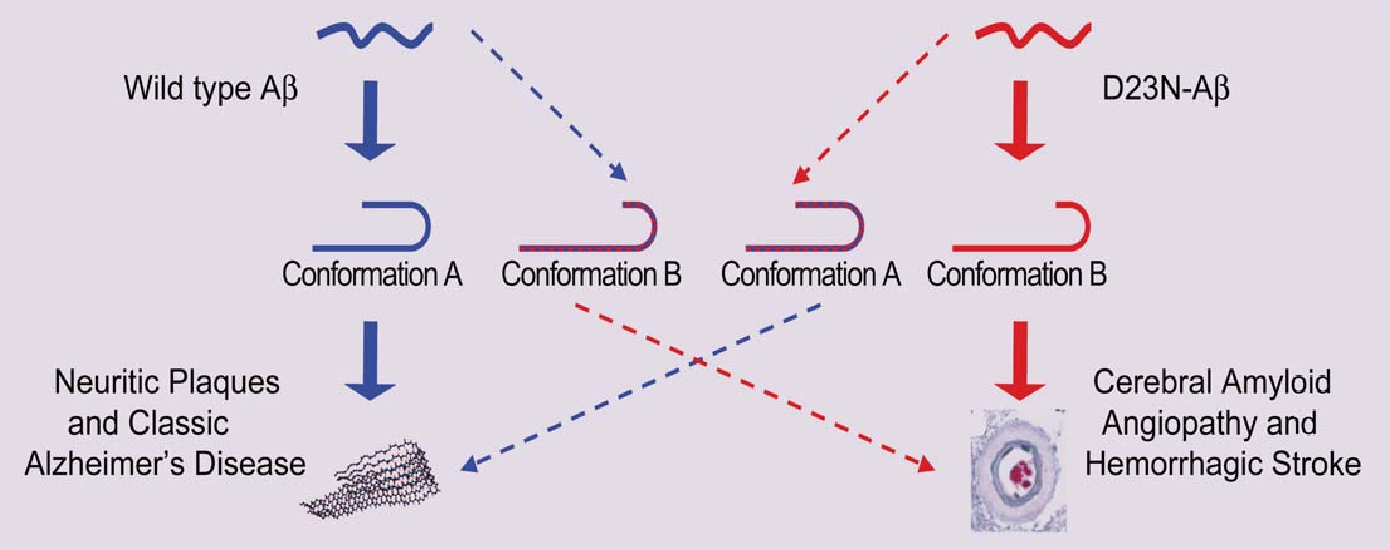
Fig. 1 Protein shape changes in wild type and mutant β-amyloids related to classic Alzheimer’s disease and hemorrhagic stroke.

For some time now, proteins known as amyloids have been implicated in the onset and advance of Alzheimer’s and other diseases, such as type 2 diabetes. One of the curious aspects of linking these proteins to diseases is that it seems to be primarily unusual protein folding and assembly that leads to disease. This is especially so in the case of Alzheimer’s and cerebral amyloid angiopathy, where mutations such as the Iowa mutant are associated with familial inheritance and early onset of the disease. Patients carrying the mutation develop neuritic plaques and large deposits of the mutant protein in cerebral blood vessels. Exactly how the protein does so much damage has been the subject of intense recent research, including these findings by researchers from The University of Chicago, the National Institutes of Health, and the Illinois Institute of Technology. With the help of the BioCAT 18-ID beamline at the APS, the team used x-ray diffraction, electron microscopy, and nuclear magnetic resonance (NMR) spectroscopy to show how the mutant and normal proteins differ with respect to folding and …

more ...

Getting to Know Cellulose

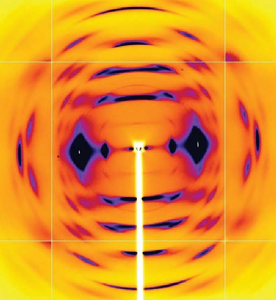
Fig. 1. Color palette figure showing x-ray data collected at the NE-CAT beamline from fiber samples of cellulose that have been converted into cellulose IIIII.

As humans continue to deplete the Earth’s supply of fossil fuels, finding new sources of energy becomes a priority. Biomass, such as cornhusks left after harvest, is one such alternative energy source. Before efficient use can be made of such materials, understanding how to break down cellulose—the fiber in human nutrition and the main component of much biomass waste—is crucial. With the help of the NE-CAT and BioCAT beamlines at the APS and the SPring-8 (Japan) beamline BL38B1, an international research team from Los Alamos National Laboratory, the University of Tokyo, and the University of Grenoble has identified important new features of cellulose structure. Their work provides important new details that could be used in designing more efficient treatments for cellulosic biomass.

Cellulose is a complicated macromolecule and only a few living things, including the microbes inhabiting the stomachs of cows and other ruminates, have figured out how to metabolize it. Yet biochemists and biophysicists have made significant progress in learning how cellulose is put together and how to …

more ...

The Power of Proteins: Prion Diseases Demystified

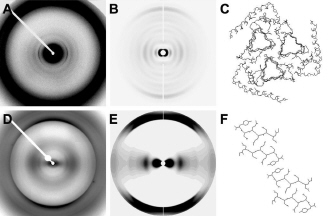
Diffraction from natural and recombinant prions: the observed x-ray diffraction pattern (A) from natural prions best fits the calculated pattern (B) from a 3-protofilament helical model (C); the observed x-ray diffraction pattern (D) from recombinant prions best fits the calculated pattern (E) from a stacked β-sheet model (F). (From H. Wille et al.,, Proc. Natl. Acad. Sci. USA 106(40), 16990 [2009], Copyright 2009 National Academy of Sciences, U.S.A.)

It is hard to believe that a single protein can be responsible for the damage inflicted by diseases such as human Creutzfeldt-Jakob and bovine spongiform encephalopathy (Mad Cow Disease). Yet the implicated protein, known as a prion and only about 200 amino acids long, can initiate and propagate a disease cycle just by changing its shape. Prions are amyloids, which are misfolded proteins now implicated in numerous diseases. Studying the prion diseases has required patience and fortitude because of disorder and insolubility of the prion samples. Aided by four U.S. Department of Energy x-ray beamlines (BioCARS 14-BM and BioCAT 18-ID at the Advanced Photon Source at Argonne, the 4-2 beamline at the Stanford Synchrotron Radiation Laboratory, and 12.3.1 at the Advanced Light Source), a …

more ...

A Closer Look at Protein Breathing

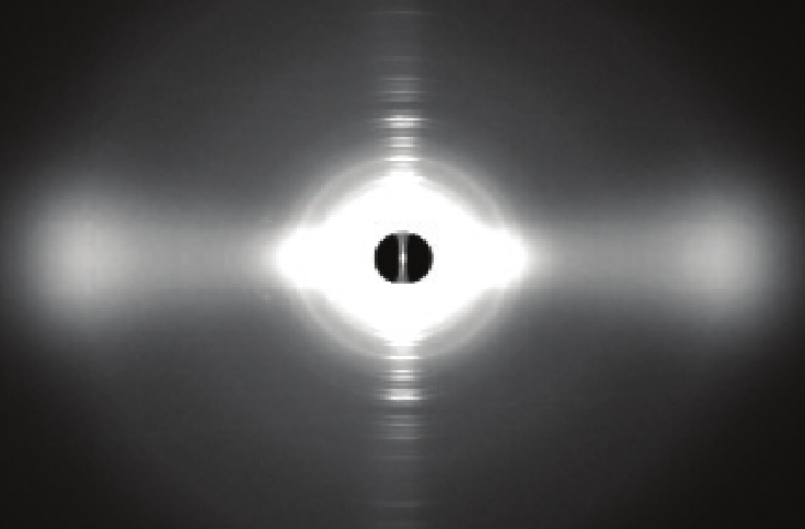
Fig. 1. A false-color scattering pattern from the protein myoglobin.

To take a static view of proteins and regard them as simple strings of amino acids that do grunt work in cells would be a mistake. Decades of biomedical research have proven that proteins are often large, complex in structure, and, as is becoming increasingly apparent, undergo sophisticated changes in space and time in order to keep cells functioning properly. Some proteins, when in solution, exhibit dramatic fluctuations in their three-dimensional structures, movement that looks like breathing. Because this movement has usually been studied in relatively dilute solutions, and not in the crowded interior of a cell, it has been difficult to know how much of the motion would actually occur in living systems. Recognizing the need for a new approach to the problem, researchers used the APS to study the breathing motions of a diverse group of five animal proteins. Their results provide badly needed modeling of protein movement in solution and data that can be used widely in biomedical applications, such as therapeutic drug design.

The researchers from Argonne National Laboratory and the Illinois Institute of Technology used computational modeling and wide-angle x-ray scattering (WAXS) experiments performed on …

more ...

Revealing the Structural Secrets of Plant Viruses

Fig. 1. Segments of the potyvirus soybean mosaic virus; several virions are also shown in cross-section. The symmetry and low-resolution structure of this virus are very similar to those of the potexvirus potato virus X, suggesting that flexible filamentous plant viruses share a common coat protein fold and approximate helical symmetry.

Viruses are extremely successful at finding ways to circumvent just about every host defense system. The secrets to this success seem to lie in their simplicity—small genomes and a metabolism that relies in part on the biochemistry of the host— and in their elegant, often breathtakingly beautiful and highly functional structures. Viral architecture, especially the coat protein structure, is intricately intertwined with successful invasion and infection of the host. Yet for many viruses, and particularly for a very large group of plant viruses, details of their structures have remained elusive. But researchers using a high-brilliance x-ray beamline at the APS have obtained important details about the structures of a soybean and a potato virus. This is good news for crop scientists concerned with finding ways to combat viral infestations.

Over the years, scientists have has invested much time and effort searching for methods to learn more about the …

more ...

New study may shed light on protein-drug interactions

Fig. 1. In this e-coli cell, the proteins (shown in blue) crowd around ribosomes (purple). These regions have a high concentration of protein, typically greater than 30 percent, which limits the ensemble of states into which the proteins can bend themselves.

Proteins, the biological molecules that are involved in virtually every action of every organism, may themselves move in surprising ways, according to a recent study from the U.S. Department of Energy’s Argonne National Laboratory that may shed new light on how proteins interact with drugs and other small molecules.

This study, which relied on the intense X-ray beams available at Argonne’s Advanced Photon Source, uses a new approach to characterize the ways in which proteins move around in solution to interact with other molecules, including drugs, metabolites or pieces of DNA.

Proteins are not static, they’re dynamic,” said Argonne biochemist Lee Makowski, who headed the project. “Part of the common conception of proteins as rigid bodies comes from the fact that we know huge amounts about protein structures but much less about how they move.”

The study of proteins had long focused almost exclusively on their structures, parts of which can resemble chains, sheets or …

more ...

Mechanisms of a Molecular Trash Disposal

Fig. 1. In this e-coli cell, the proteins (shown in blue) crowd around ribosomes (purple). These regions have a high concentration of protein, typically greater than 30 percent, which limits the ensemble of states into which the proteins can bend themselves.

The ubiquitin proteasome system (UPS) is part of the waste management system of a cell. Proteins that are destined to have limited life spans in the cell are identified and sorted for destruction. This is accomplished by labeling the proteins with the molecule ubiquitin, which is present in all eukaryotes. Once tagged, the proteins are degraded by a complex machine known as the proteasome. The UPS is not just a molecular wood chipper; it performs its function with specificity that is provided by the class of ubiquitination enzymes called E3 ligases. The E3 ligases identify proteins to be degraded through domains called F-boxes, but the mechanism of enzymatic transfer of ubiquitin to the doomed protein is still not completely understood and may be facilitated by the dimerization of another domain, known as the D-domain. In studies carried out at the NE-CAT 8-BM-B, BioCars 14-BM-C, BioCAT 18-ID-D, and SBC-CAT 19-BM-D …

more ...

Muscle-Fiber Research Expands

Fig. 1. The upper panel (a) shows the M3 x-ray reflection from an active single muscle fiber at constant length (L0) recorded at APS. The reflection is split into two peaks of roughly equal intensity by interference between the two arrays of myosin motors shown in red in the cartoon representation of the muscle sarcomere (b). When the load on the muscle fiber is decreased (c) it shortens, reaching a steady speed (at L4) that depends on the load. The M3 reflection recorded at L4 (d) has interference peaks of markedly different intensities (middle panel), and this signals motion of actin-attached myosin motors towards the midpoint of the sarcomere combined with some motor detachment (e).

It has been known that muscle fibers shorten slowly under heavier loads and faster under lighter loads. Now, researchers from the Università degli Studi di Firenze, Università di Roma, Dexela Ltd., the Illinois Institute of Technology, and King’s College London, using the BioCAT 18-IDD beamline at the APS have discovered the molecular basis of this fundamental property of muscle function. Their work was the cover article for the issue of Cell magazine in which it was published. Understanding exactly how muscle fibers work …

more ...

Deconstructing Heart Muscle

Fig. 1. . X-ray diffraction patterns of normal mouse heart muscle (WT, bottom) and heart muscle from mice lacking cardiac myosin-binding protein-C (KO, top). Compared with WT muscle, the KO muscle matter is shifted toward its thin actin filaments (represented by the size of the outermost black dots in both images), as opposed to its thick myosin filaments (inner dots), implying that cardiac myosinbinding protein-C helps keep myosin filaments more tightly wound than they would be otherwise.

The stars of muscle contraction—be it the flex of a bicep or the throb of the heart—are microscopic fibers spun from the proteins actin and myosin. To play their parts properly, these proteins need help from an additional, less well-understood molecule known as myosin-binding protein-C. Thanks to data collected at BioCAT beamline 18-ID-D at the APS, researchers from the University of Wisconsin Medical School and the Illinois Institute of Technology have gleaned insights into this protein’s contribution to a smoothly beating heart.

Heart muscle fibers, like those of skeletal muscle, consist of alternating sets of parallel filaments. Thick filaments, which are bundles of myosin molecules, overlap at each end with thin filaments composed of small actin molecules. Connecting the …

more ...