Targeting Cancer at the Level of DNA Expression

Small-angle x-ray scattering-refined molecular models of oncogenic promoter G4s. Shown are best-fitting atomic models of the various higher order G4 promoter sequences from c-Myc, k-Ras, and c-Kit promoters displayed with transparent molecular surface representations.

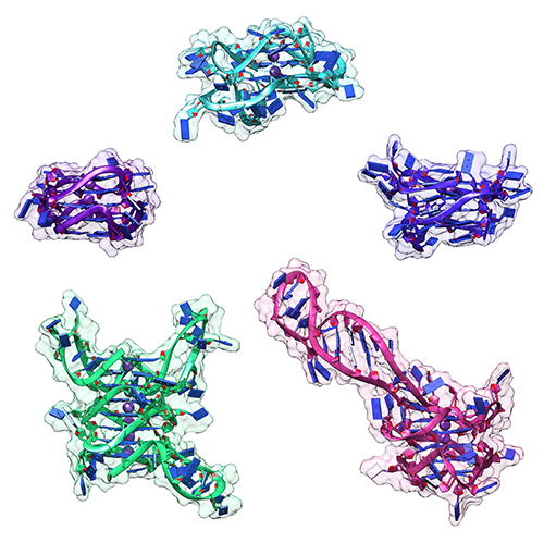
The last 20 years have brought a revolution in targeted therapies for cancer. Small-molecule inhibitors and monoclonal antibodies that target a specific aberrant protein in tumors have provided cancer patients with treatments that are associated with fewer side effects and longer survival than conventional chemotherapy. This has been, in large part, the result of intensive research into the role of oncogenes in cancer development. Oncogenes are normal cellular genes that have become mutated in such a way that they aberrantly promote the uncontrolled cell growth seen in cancer. They are often proteins involved in growth control or activation of cellular signaling; inhibiting these mutated proteins has proven to be effective in stopping the growth of many cancers. Research by a team from the Brown Cancer Center at the University of Louisville in Kentucky using the U.S. Department of Energy’s Advanced Photon Source (APS) and published in the journal Nucleic Acids Research promises to extend these treatment possibilities to control these oncogenes at the gene …

more ...

Understanding the Structural Implications of Genetic Mutations in Heart-Muscle Disease

Mechanism of action for HCM-D166V and DCM-D94A mutations. The HCM-D166V model disrupts the SRX state and promotes the SRX-to-DRX transition increasing the number of DRX heads and leading to hypercontractile behavior. The DCM-D94A model stabilizes the SRX state yielding fewer heads available for contraction and leading to clinical hypocontractility. Abbreviations: ELC, myosin essential light chain; RLC, regulatory light chain; DRX, disordered relaxed; SRX, super-relaxed.

Cardiomyopathies are diseases of the heart muscle in which the muscle of the pumping chamber (ventricle) can become enlarged (dilated cardiomyopathy; DCM) or thickened (hypertrophic cardiomyopathy; HCM), potentially leading to heart failure. There are currently no effective treatments but the disease often has a genetic component related to mutations in the heart muscle proteins that are involved in muscle contraction, so some researchers have focused their therapeutic development efforts on correcting these muscle contraction problems based on the structural basis of the defect. A recent study from a team of researchers using the U.S. Department of Energy’s Advanced Photon Source (APS) employed humanized mouse models expressing mutations observed in patients with HCM and DCM to evaluate the structure-function relationships and the changes observed in cardiac muscle contraction with …

more ...

New Resource for the Muscle Diffraction Community

Three possible conformations of an intrinsically disordered protein: collapsed (purple), expanded (gold) and a combination of collapsed and expanded (red). Image created by Kristina Davis, University of Notre Dame.
BioCAT staff have just published a review article, Ma & Irving, 2022 Int. J. Mol. Sci. 2022, 23(6), 3052, on the use of small angle X-ray fiber diffraction for studying skeletal and cardiac muscle disease. The article consists of a guided tour of the various diffraction features that can be used to extract specific pieces of information that can be used to provide insights into the structural basis of pathology. The article also contains a comprehensive review of the literature reporting diffraction studies of muscle that illustrates how small angle fiber diffraction has increased our understanding of specific muscle diseases such as hypertrophic cardiomyopathy, dilated cardiomyopathy, and nemaline myopathy.
more ...

What Bacterial Pathogens Can Teach Us about Protein Folding

Three possible conformations of an intrinsically disordered protein: collapsed (purple), expanded (gold) and a combination of collapsed and expanded (red). Image created by Kristina Davis, University of Notre Dame.

Protein folding is one of the fascinating unanswered questions in biology. How does an amino acid sequence that is unfolded when it leaves the ribosome manage to fold properly into a highly ordered, lightning-fast enzyme or sturdy structural protein? Why don’t all the proteins in the cell instead just stick to each other, aggregating into a big mess? A unique model system in bacteria may hold some of the answers to these questions. The system involves the study of what are termed autotransporter proteins, which pathogenic bacteria secrete as virulence factors for infection. These proteins are synthesized in the bacterial cytoplasm and cross one membrane into the bacterial periplasm. Autotransporter proteins then remain in an unfolded state in the periplasm until they pass through the outer bacterial membrane, folding properly along the way. This highly specialized protein folding process has attracted the attention of a team of researchers who have used this bacterial system as a model to determine what allows these unique proteins to maintain their disordered state in …

more ...

Relaxation at the Molecular Level

Two-dimensional x-ray diffraction patterns from resting (left) and contracting (right) mouse soleus muscle and the corresponding myosin structures. The differences in myosin structures (scallop myosin) during resting (left) and contracting (right) can be detected by the x-ray patterns. Pattern changes were tracked in live muscle in a time resolved manner.

The molecular interactions between the proteins myosin and actin that generate force during muscle contraction are some of the most well-studied molecular interactions in biology. However, there are some congenital skeletal muscle disorders and types of heart failure where relaxation of the muscle, rather than the force generation part of the cycle, appears to be the problem, and there are currently no available treatments that affect relaxation specifically. A more detailed understanding of the dynamics of the relaxation process could help in the development of treatments that maintain or increase force generation while repairing defects in relaxation. Recent work conducted at BioCAT used a unique transgenic mouse model, time-resolved small-angle x-ray diffraction, and molecular dynamics simulations to discover more about how myosin and actin interact during skeletal muscle relaxation. This research, published in the Journal of Physiology, demonstrates that this type of small-angle x-ray analysis may be of great value …

more ...

Understanding the Physiology of the Human Heart through the Study of Tarantula Muscles

The human heart is a remarkable feat of evolutionary engineering. Beating about 100,000 times per day and pumping nearly 2,000 gallons of blood through an interconnected series of veins, arteries, and capillaries that spans a distance greater than 60,000 miles, the heart is the most important muscle in the human body. Yet, heart disease remains the number one cause of death in the world, demonstrating the need for more research in heart physiology. Now a research team has found an unlikely source of inspiration for understanding how the human heart works and how we might design better drugs for conditions like hypertrophic cardiomyopathy: tarantulas. The source of nightmares for arachnophobes and the household pets for arachnophiles are inspiring researchers to take new approaches to understanding diseases that alter how heart muscle cells contract and relax. But, before getting to the human heart, there is more to learn about the physiology of tarantula muscles. The researchers set out to understand how contractions in tarantula muscle cells are activated and why are muscle twitches that follow a sustained muscle contraction (post-tetanic) more forceful than those that don’t (pre-tetanic). Their results provide evidence that phosphorylation, the chemical addition of …

more ...

Key Insights into an Inherited Muscle Disease

Compound nebulin mutations cause changes in thin filament structure.

The gene NEB encodes for the skeletal muscle protein nebulin. Mutations in NEB cause the disease nemaline myopathy, which is one of the more common inherited myopathies. Patients with this muscle disorder have muscle weakness in multiple different parts of their body and can also experience difficulties with feeding or breathing. Currently, there is no cure for nemaline myopathy and treatment options are limited. Therefore, there is a need to better understand this disease and design new therapeutics that can improve patient quality of life. A team of researchers from the University of Arizona working to provide new insights into the pathogenesis of this skeletal muscle disorder, report a new mouse model of nemaline myopathy that exhibits similar symptoms to those identified in human patients. An important part of this work utilized x-ray diffraction data collected at the APS. The diffraction data provide new insights into how mutations of NEB alter the molecular structure of skeletal muscles. The findings from the study are highly impactful because they significantly increase our understanding of how mutations in the gene NEB cause nemaline myopathy. Importantly, the new mouse model of this disease can be …

more ...

Structure-Function Studies Elucidate GPCR-Independent Regulation of G-proteins

Guanine nucleotide binding proteins popularly known as G-proteins, involved in a variety of cellular signal transduction pathways are heterotrimeric proteins consisting of α, β, and γ subunits. Ric8A is known to be both a chaperone for the assembly of the α-subunit of G-proteins, and a Guanine nucleotide Exchange Factor (GEF). McClelland et al., have conducted a detailed structural analysis on the complex between Ric8A and Gαi1 using cryoEM, X-ray crystallography, and SAXS. Constructs of Ric8A and Gαi1 optimized for structure determination and to reduce conformational heterogeneity were used to assemble the Ric8A-Gαi1 complex. They were able to determine the interface between the two proteins which consists of three separate surface contacts which essentially stabilize Gα in its nucleotide-free state. Furthermore, it was found that a specific Casein Kinase mediated phosphorylation of Ric8A stimulated the GEF activity by structurally stabilizing the Ric8A-Gα interface. Ric8A binding to the disrupted Guanine nucleotide binding site on Gα was determined to be critical for the GEF function as point mutants at residues of Ric8A involved in this interaction compromised the GEF activity. A significant amount of re-organization of a particular helical domain in Gα relative to the GTPase domain is involved in providing a means …

more ...

Uncovering Unique Structural Features in Protein Regions Associated with ALS

Single chain of the model (top), microscopy of liquid-liquid pase separation (middle), and images from phase separating simulations (bottom).

Many of us are familiar with mad cow disease–the neurodegenerative disease caused by prions. Although they have a similar name, the less familiar prion- like domains (PLDs) refer to something different–unique, low-complexity regions of proteins that are capable of regulating gene expression and affecting important cellular processes. Prion-like domains have become a topic of interest because of their connection with a variety of debilitating brain diseases, such as amyotrophic lateral sclerosis (ALS) and frontotemporal dementia. In fact, mutations in PLDs of some genes have been shown to cause neurodegenerative disease. For example, mutations in PLDs of the genes hnRNPA2B1 and hnRNPA1 can cause the neurodegenerative disorders ALS and multisystem proteinopathy. A recent study using data obtained at BioCAT completed a comprehensive biophysical investigation of PLDs in the protein hnRNPA1 to uncover the major behavioral and structural features of these domains. This meaningful work may lead to discoveries that can help individuals living with such neurodegenerative diseases.

Amyotrophic lateral sclerosis is a devastating disease of the nervous system that affects the brain and spinal cord. Patients often present with symptoms …

more ...

Sarcomere Structure and Nemaline Myopathy

NEM6 mutations cause changes in thin filament structure.

Nemaline myopathy (NM) is one of the most common congenital non-dystrophic myopathies and is characterized by severe hypotonia, muscle weakness, feeding difficulties, respiratory failure, and the presence of nemaline bodies (rods) in skeletal muscle biopsies. One form of nemaline myopathy is caused by mutations in the KBTBD13 (NEM6) gene. In addition to weakness, NEM6 patients have slow muscle relaxation, compromising contractility and daily-life activities. The role of KBTBD13 in muscle is unknown, and the p athomechanism underlying NEM6 is undetermined. A combination of transcranial magnetic stimulation-induced muscle relaxation, muscle fiber- and sarcomere-contractility assays, super-resolution microscopy, and low angle X-ray diffraction on the BioCAT Beamline 18ID revealed that the impaired muscle relaxation kinetics in NEM6 patients are caused by structural changes in the thin filament, a sarcomeric microstructure. Using homology modeling, binding- and contractility assays with recombinant KBTBD13, novel Kbtbd13-knockout and Kbtbd13R408C-knockin mouse models and a transgenic zebrafish model the authors discovered that KBTBD13 binds to actin – a major constituent of the thin filament - and that mutations in KBTBD13 cause structural changes impairing muscle relaxation kinetics. The authors propose that this actin-based impaired relaxation is central to NEM6 pathology.

See: Josine M. de …

more ...