NUCLEOTIDE-INDUCED ASYMMETRY WITHIN ATPASE ACTIVATOR RING DRIVES σ54-RNAP INTERACTION AND ATP HYDROLYSIS

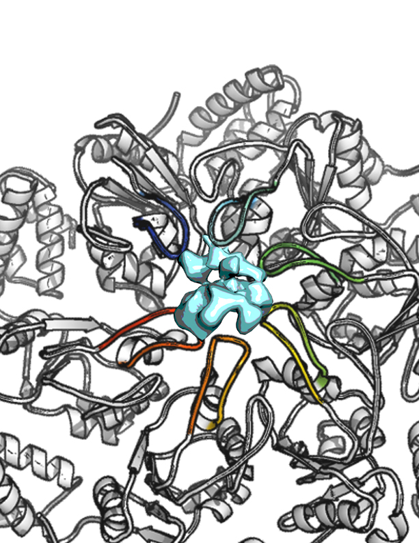
Living creatures use ATP as the “universal energy currency”. ATP-ases are assemblies of molecules that break down ATP into smaller molecules using the energy released to power myriad biological reactions. Molecular motors are ATP-ases that convert this chemical energy into mechanical work on other molecules. The AAA+ ATPases are examples of such molecular machines that perform mechanical work to remodel nearly every type of macromolecule, in cells from all kingdoms of life. A long-standing, largely unanswered question about the functional mechanism of the AAA+ ATPases is how do the rings of chemically identical subunits that make up these assemblies interact with their target macromolecules? The authors address this question by studying Enhancer Binding Proteins (bEBPs) in bacteria, AAA+ ATPases that remodel the σ54-form of RNA polymerase (Eσ54) that is present in complexes with promoter DNA. This remodeling or shape transformation is essential to allow transcription and subsequent expression of genes that allow for nutrient acquisition, complex developmental programs, and virulence as pathogens.

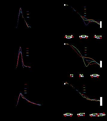
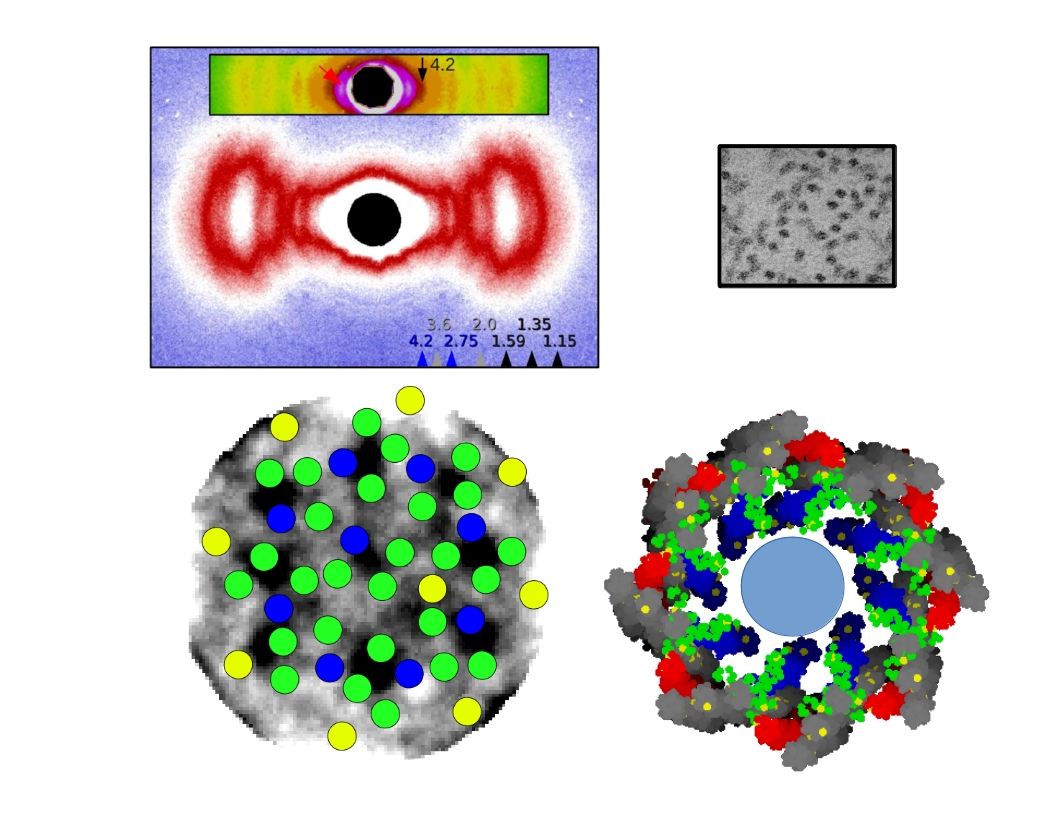
In the current work the authors used isothermal calorimetry (ITC), crystallography and 3D reconstruction from EM single particles along with time-resolved and static small angle X-ray scattering (TR-SAXS and SAXS, respectively) at BioCAT to monitor …

more ...

Conformational states and recognition of amyloidogenic peptides of human Insulin-degrading enzyme

SAXS analysis of IDE. Pair distribution functions and scattering curves of WT IDE and IDE R767A (C and D), and IDE S132C/E817C (E and F). Curve fitting is based on atomic models using the program CRYSOL (single model) or OLIGOMER (mixture). D2/D3, D2/D3 pivot; D1/D4, D1/D4 pivot; C, closed state; M, monomer; D, dimer; T, tetramer. The diagrams and ratios shown below the scattering profiles represent the distribution of mixture that could best fit the SAXS data.

Proteins in living organisms face acute and chronic challenges to their integrity, which require proteostatic processes to protect their functions. Proper protein function is ensured through protein turnover through a balance between synthesis and proteolysis. Amyloidogenic peptides, such as amyloid β (Aβ) and amylin, present a major challenge to proteostasis, because they can form toxic aggregates that impair diverse physiological functions and contribute to human diseases. Insulin-degrading enzyme (IDE), a Zn2+-metalloprotease, prefers to degrade amyloidogenic peptides to prevent the formation of amyloid fibrils. Thus, IDE retards the progression of Alzheimer’s disease.

IDE possesses an enclosed catalytic chamber that engulfs and degrades its peptide substrates; however, the molecular mechanism of IDE function, including substrate access to …

more ...

Molecular packing and fibrillar structure of type II collagen

Type II collagen is the principal extracellular matrix (ECM) component of mammalian cartilage. It has been shown that lamprey (a cartilagenus fish) notochord collagen fibrils are indistinguishable from human cartilage fibrils, their differences being minor amino acid sequences and in the specific arrangement of the fibrils to build their respective ECM’s. Recently, the Orgel group demonstrated that a human anti-biglycan antibody had the capacity to ‘deconstruct’ lamprey notochord type II collagen fibril bundles to give a pure source of ‘thin-fibril’ collagen fibrils that are primarily composed of type II collagen. This procedure provides both a possible explanation for the initiation of Rheumatoid arthritis and a means of studying the structural arrangement of tissue.

This work comprised two principal methods: 1) Using highly focused microbeam at BioCAT to locate the most crystalline portions of a notochord sample, and collecting data from this region over extended periods of time (>15 minutes) using a cryojet. 2) Analysis of TEM data from fibril cross-sections to identity the arrangement of collagen microfibrils within the thin-fibrils. Using the first method, the Orgel group determined the quasi-hexagonal packing of the notchord tissue (previously thought to be tetragonal due to limitations in data available) and the 2D …

more ...

Non-enzymatic decomposition of collagen fibers by a biglycan antibody and a plausible mechanism for rheumatoid arthritis

Type II collagen fibrils decomposed into their basic aggregates (viewed via X-ray diffraction and TEM). Some parts of the antibody treated samples maintain a loose alignment of the thin-fibrils allowing them to be analyzed with small angle X-ray diffraction (A), and insert B. An 11 and 4.5 nm packing function are apparent, which appear to correspond to the approximate diameter of the thin-fibrils (insert of C) and microfibrils (D). Native thick fibrils are shown in C as a comparison to the decomposition product (thin-fibrils).

Rheumatoid arthritis (RA) is a systemic autoimmune inflammatory and destructive joint disorder that affects tens of millions of people worldwide. Normal healthy joints maintain a balance between the synthesis of extracellular matrix (ECM) molecules and the proteolytic degradation of damaged ones. In the case of RA, this balance is shifted toward matrix destruction due to increased production of cleavage enzymes and the presence of (autoimmune) immunoglobulins resulting from an inflammation induced immune response.

In this study, the authors demonstrate that a polyclonal antibody against the proteoglycan biglycan (BG) causes tissue destruction that may be analogous to that of RA affected tissues. The effect of the antibody is more potent than various harsh chemical and/or …

more ...

The cross-bridge spring: cool muscles store elastic energy

Example negative work-loop at 25ºC and positive work-loop at 35ºC. The red dot indicates the point of muscle activation and the black dots represent times when diffraction images were collected throughout the contraction cycle. Diffraction images from the time point directly following muscle stimulation highlight the temperature dependent variation in the lattice structure.

The Hawkmoth Manduca sexta is an emerging model system for a wide range of studies in integrative biology. The flight muscles are particularly interesting in that, unlike most insect flight muscle, but like vertebrate skeletal and cardiac muscles, they are a synchronous muscle where each stimulus generates one muscle twitch. The length tension curve also shows intriguing similarities to mammalian cardiac muscle even though the sarcomere structure is known to be quite different. Another property of the muscle is that the dorsal-most region of the flight muscle is ca. 5 degrees C cooler than the ventral muscle closer to the midline to the body. (Such spatial temperature gradients are also likely to occur in large muscles in mammals but this has not been well investigated). In Manduca flight muscle these spatial gradients lead to a spatial variation in power production spanning from positive to negative values across …

more ...

A New Phase in Cellular Communication

Multivalency drives phase separation and probably drives a sol–gel transition in the droplet phase. a, Phase diagrams of multivalent SH3 and PRM proteins. The concentrations are in terms of the modules. The red circles indicate phase separation, and the blue circles indicate no phase separation. b, The Rg values determined from SAXS data that were collected during titrations of PRM proteins into SH35. Closed circles indicate the absence of phase separation; open circles indicate data collected on the supernatant phase,which was separated from the droplets by centrifugation. The titrations used PRM4 (orange), PRM2 (blue), PRM1 (green) and PRM(H)1 (red). The error bars represent the s.d. calculated fromfive to ten independent measurements of intensity versus scattering angle (q). c, The intensity autocorrelation curve of light scattered at 90u from the pooled droplet phase of SH35 plus PRM(NWASP) 8. t, the relaxation time constant of the most rapidly decaying phase. d, Cryo-electron microscopy image of a droplet formed by SH35 plus PRM5 (identical image, left and right).

In many biological processes, various substances undergo phase transitions, where they are transformed from one state (solid, liquid, or gas) to another. Wiskott-Aldrich Syndrome Proteins (WASP) function as intracellular …

more ...

RNA Folding – How A Little Cooperation Goes A Long Way

Cooperative Folding of the Azoarcus Group I Ribozyme (A) Compact, native-like intermediates (IC) form in low Mg2+ (Perez-Salas et al., 2004; Rangan et al., 2003) and are detected by SAXS or native PAGE. Formation of the native structure (N) is reported by ribozyme activityand the solvent accessibility of the RNA backbone. See also Figure S1. (B and C) Tertiary interaction motifs indicated by red dots were perturbed by single-base substitutions: loop L2, A25U; joining region J2/3, A39U; paired region P6, A97U; TH, G125A; loop L9, A190U (see Table S1). J8/7, cyan ribbon. Cooperative interactions are indicated by red (IC) or blue (N) arrowheads (positive, pointed; negative, flat). Thickness indicates relative strength. (C) Ribbon drawn with PyMOL; 1u6b (Adams et al., 2004b).

The nucleic acid RNA plays an important role in protein synthesis in cells. However, noncoding RNAs also exist that are not converted into proteins, but still play important roles in many biological processes. RNA molecules aggregate into complex tertiary structures, producing globular forms stabilized by various interactions. Proteins, ligands, and other RNA molecules recognize tertiary folded RNAs and result in the biochemical pathways that affect all aspects of cellular metabolism. Using small-angle X-ray scattering (SAXS) at the …

more ...

The Molecular Mechanism of Stretch Activation in Insect Muscle

X-ray pattern from contracting flight muscle. Top: Match-mismatch of crossbridge origins with actin target zones. Bottom: Thick filament twisting bring myosin crossbridges closer to actin binding sites (“target zones”). Pink = target zones; red = myosin heads. Inset: Lethocerus indicus.

Flying insects are among the most successful species on our planet. Flight is very metabolically demanding and many insects have found a clever way to reduce energy costs in their flight muscles by employing a process called “stretch activation, which has been recognized since the 1960s as an interesting and physiologically important phenomenon, but a mechanistic explanation has been elusive. Now, research at the Biophysics Collaborative Access Team x-ray facility at the U.S. Department of Energy’s Advanced Photon Source provides another, important step toward a full explanation of stretch activation, which also plays an important role in mammalian cardiac expansion and contraction.

How stretch activation works in the heart is unknown. As contractions propagate through the heart, the contraction of one piece of muscle tissue stretches adjacent muscle, thereby activating it. The end result is a very strong contraction at the end of systole aiding cardiac ejection. Heart muscle is much less organized structurally than insect muscle and is thus …

more ...

An Understanding of Elastin’s Properties Springs Forth

Top: Ab initio shapes of full-length tropoelastin calculated from solution SAXS or SANS data. The filtered average shapes of 20 individual SAXS (red) and SANS (yellow) simulations are shown as a surface representation. An overlay of the models from the two scattering methods is also shown. The proposed locations of the N-terminus, the spur region containing exons 20-24 and the C-terminus are indicated. Scale bar is 5 nm. Middle: SAXS analysis of overlapping fragments of human tropoelastin. Ab initio models were calculated from SAXS data for tropoelastin constructs 2-18 (blue), 2-25 (brown) and full-length (red). An overlay of the two N-terminal fragments shows a conserved linear region. Scale bar is 5 nm. Bottom: Head-to-tail model for elastin assembly. A) Juxtaposed domains 19 and 25 on one tropoelastin molecule and domain 10 on an adjacent monomer would allow the formation of a three-way desmosine cross-link found in vivo. B) Tandem assembly of tropoelastin monomers displaying n-mer propagation as an outcome of covalently bonded molecules.

It’s not stretching the truth to say that flexibility is an important and desirable human physiological trait. We owe our flexibility to a protein called elastin, and elastin derives its properties from a building-block molecule called …

more ...

How Dinosaurs Put Proteins into Long-Term Storage

X-ray diffraction model of the rat collagen microfibril showing the location of sites for fibronectin (Fn), decorin, and integrin binding, matrix metalloproteinase (MMP) cleavage, and the location of putative cell and matrix interaction domains. Dinosaur peptide locations are shown in red, green, and blue. Image originally published in San Antonio et al., PLoS ONE 6(6), e20381 June 2011).

How does one prove that the protein isolated from a 68-million-year-old dinosaur bone is not a contamination from the intervening millenia or from the lab? This is the task of a research team who say they have isolated peptides of the common structural protein, collagen, from bones of Tyrannosaurus rex and Brachylophosauraus canadensis. Although the team had previously presented multiple lines of evidence supporting the veracity of the find, the fact that the age of the peptides far exceeds any previous predictions of how long a protein could resist degradation has generated controversy. In their current work, the researchers used x-ray diffraction data collected utilizing the BioCAT 18-ID x-ray beamline at the U.S. Department of Energy Office of Science’s Advanced Photon Source at Argonne National Laboratory to generate a model of collagen structure on which to overlay the …

more ...