Increased Brain Iron Coincides with Early Plaque Formation in a Mouse Model of Alzheimer’s Disease

Spatial distribution of Fe, Cu, and Zn in the hippocampus of PSAPP and CNT mice measured using XFM. (A) H&E stained hippocampal brain section from a PSAPP mouse. XFM images of (B) Fe, (C) Zn, and (D) Cu in a serial tissue section. Units are mM. Scale bar = 300 μm.

Early and correct diagnosis of Alzheimer’s disease (AD) is important for reasons that go beyond correct diagnosis and treatment of symptoms. These reasons include more time to make critical life decisions, planning for future care, and maximizing the safety of the person with Alzheimer’s disease and their family. New scientific results relevant to the diagnosis and treatment of AD have been obtained by researchers utilizing the U. S. Department of Energy’s Advanced Photon Source (APS) at Argonne National Laboratory and National Synchrotron Light Source (NSLS) at Brookhaven National Laboratory, and published in the journal NeuroImage. This work points to the use of elevated brain iron content, already observed in late-stage AD, as a potential tool for early diagnosis. Since the disease is usually diagnosed only in later stages after cognitive symptoms appear and treatment may not be effective, a method for early detection would be a …
more ...

The Molecular Mechanism of Stretch Activation in Insect Muscle

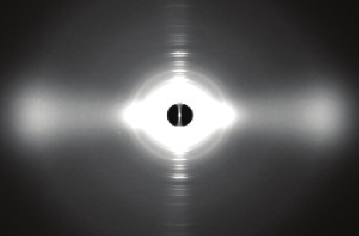
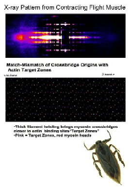
X-ray pattern from contracting flight muscle. top: Match-mismatch of crossbridge origins with actin target zones. bottom: Thick filament twisting bring myosin crossbridges closer to actin binding sites (“target zones”). Pink = target zones; red = myosin heads. Intruder at bottom: Lethocerus indicus.

Flying insects are among the most successful species on our planet. Flight is very metabolically demanding and many insects have found a clever way to reduce energy costs in their flight muscles by employing a process called “stretch activation,” whereby nervous stimulation is just enough to maintain a constant low level of calcium and the muscles are “turned on” when they are stretched by antagonistic muscles. Stretch activation has been recognized since the 1960s as an interesting and physiologically important phenomenon, but a mechanistic explanation has been elusive. Now, research at the Biophysics Collaborative Access Team (BioCAT) synchrotron x-ray facility at the U.S. Department of Energy’s Advanced Photon Source (APS) at Argonne provides another, important step toward a full explanation of stretch activation, which also plays an important role in mammalian cardiac expansion and contraction.

How stretch activation works in the heart is unknown. As contractions propagate through the heart, the contraction of one piece of muscle tissue …

more ...

At the Crossroads of Chromosomes

Structure of the centromere histone complex containing two chains of CENP-A (red) and two copies of its close binding partner, histone H4 (blue). (Image: Ben E. Black, University of Pennsylvania School of Medicine)

On average, one hundred billion cells in the human body divide over the course of a day. Most of the time the body gets it right but sometimes problems in cell replication can lead to abnormalities in chromosomes—-resulting in many types of disorders from cancer to Down syndrome. Now, researchers from the University of Pennsylvania School of Medicine (UPSM) have defined the structure of a key molecule that plays a central role in how DNA is duplicated and then moved correctly and equally into two daughter cells to produce two exact copies of the mother cell. Without this molecule, entire chromosomes could be lost during cell division, so this work is a major advance in understanding the molecules driving human genetic inheritance. Two U.S. Department of Energy x-ray light sources, including the Advanced Photon Source (APS) at Argonne National Laboratory, were important tools for the researchers carrying out this study.

The UPSM researchers report, in the September 16, 2010 issue of Nature, the structure …

more ...

Packing It In: A New Look at Collagen Fibers

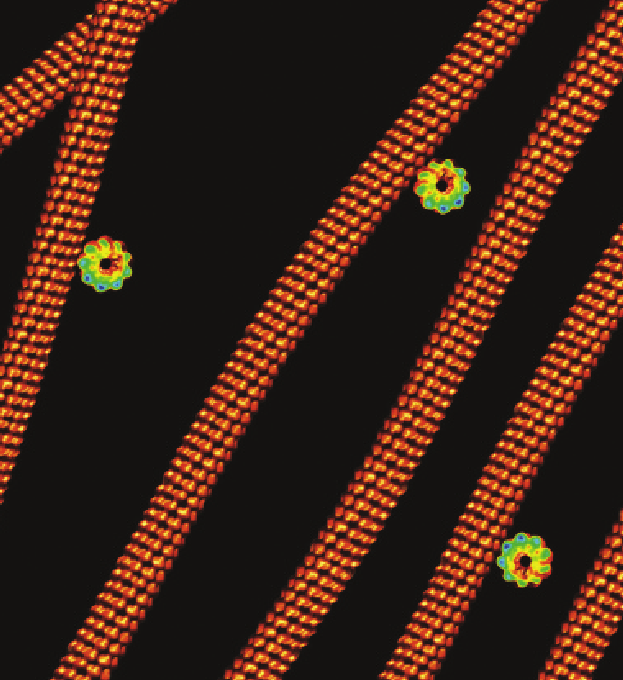
Fig. 1. Left: Medium-wide angle X-ray diffraction pattern of collagen type II fibrils from lamprey notochord. Right: 15—20 Å resolution, as seen in the meridional reflections series. (Adapted from Antipova and Orgel, J. Biol. Chem. 285(10), 7087 [March 2010] Background image of collagen fibers courtesy of Prof. Andrew Notebaert, Indiana University Bloomington. Source: Course A215, “Undergraduate Anatomy.” ©2008, The Trustees of Indiana University)

Nature uses collagen everywhere in constructing multicellular animals. There are at least 20 types of collagen, but 80-90% of the collagen in the body consists of types I, II, and III. Collagen type I is used to form skin, tendon, vascular, ligature, organs, bone, dentin, and interstitial tissues. Collagen type II makes up 50% of all cartilage protein, and is essential in normal formation of such structures as cartilage, the vitreous humor of the eye (the clear gel that fills the space between the lens and the retina of the eyeball of humans and other vertebrates), bones, and teeth. To create these structures, collagen molecules are positioned in arrays called fibrils, producing what are known as the D-periodic fibrillar collagens. Though previous work has given some idea of what the D-periodic structure looks like, technical …

more ...

The Push and Pull of Plant Viruses

Fig. 1. A new method (G2G, “Global measurements to Global structure”) has been developed for structure determination of large RNAs in solution using residual dipolar coupling (RDC) from NMR measurements, represented by the blue and red contour linked by black lines with residue labels; and SAXS/WAXS measurements, represented by gray co-centered circular rings in the background. The black “wave” is the RDC-structural periodicity correlation curve that was used to extract the orientation of the RNA duplexes. At the lower left is a two-dimensional (2-D) drawing of the topology of the 102-nt RNA. The low-center model is a rendering of the 2-D drawing of the topology in three-dimensional (3-D) space; at the lower right is the refined 3-D ensemble of the 102-nt RNA structures that were restrained with RDC and SAXS data. (For a detailed description of the G2G method, please see J. Mol. Biol. 393, 717 (2009). Cornfield photo courtesy of Sam Mugraby, Photos8.com, www.photos8.com.)

New insights into the way a simple-seeming plant virus, the turnip crinkle virus (TCV), goes about replicating in infected cells have been obtained using solution nuclear magnetic resonance spectroscopy (NMR) and small/wide angle x-ray scattering (SAXS/WAXS) studies with …

more ...

Protein Assembly and Disease

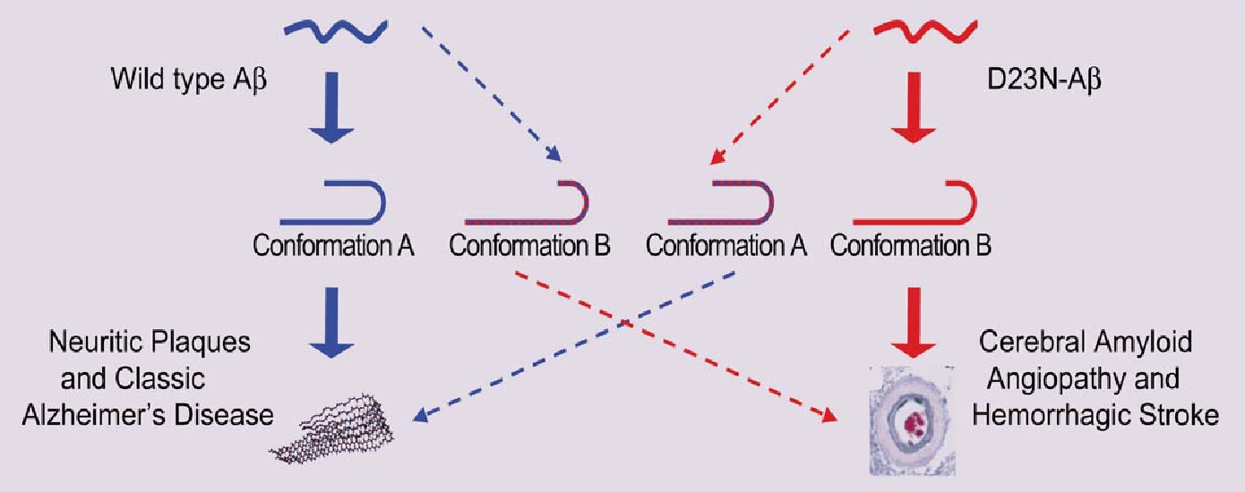
Fig. 1 Protein shape changes in wild type and mutant β-amyloids related to classic Alzheimer’s disease and hemorrhagic stroke.

For some time now, proteins known as amyloids have been implicated in the onset and advance of Alzheimer’s and other diseases, such as type 2 diabetes. One of the curious aspects of linking these proteins to diseases is that it seems to be primarily unusual protein folding and assembly that leads to disease. This is especially so in the case of Alzheimer’s and cerebral amyloid angiopathy, where mutations such as the Iowa mutant are associated with familial inheritance and early onset of the disease. Patients carrying the mutation develop neuritic plaques and large deposits of the mutant protein in cerebral blood vessels. Exactly how the protein does so much damage has been the subject of intense recent research, including these findings by researchers from The University of Chicago, the National Institutes of Health, and the Illinois Institute of Technology. With the help of the BioCAT 18-ID beamline at the APS, the team used x-ray diffraction, electron microscopy, and nuclear magnetic resonance (NMR) spectroscopy to show how the mutant and normal proteins differ with respect to folding and …

more ...

Getting to Know Cellulose

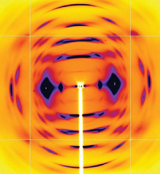
Fig. 1. Color palette figure showing x-ray data collected at the NE-CAT beamline from fiber samples of cellulose that have been converted into cellulose IIIII.

As humans continue to deplete the Earth’s supply of fossil fuels, finding new sources of energy becomes a priority. Biomass, such as cornhusks left after harvest, is one such alternative energy source. Before efficient use can be made of such materials, understanding how to break down cellulose—the fiber in human nutrition and the main component of much biomass waste—is crucial. With the help of the NE-CAT and BioCAT beamlines at the APS and the SPring-8 (Japan) beamline BL38B1, an international research team from Los Alamos National Laboratory, the University of Tokyo, and the University of Grenoble has identified important new features of cellulose structure. Their work provides important new details that could be used in designing more efficient treatments for cellulosic biomass.

Cellulose is a complicated macromolecule and only a few living things, including the microbes inhabiting the stomachs of cows and other ruminates, have figured out how to metabolize it. Yet biochemists and biophysicists have made significant progress in learning how cellulose is put together and how to …

more ...

The Power of Proteins: Prion Diseases Demystified

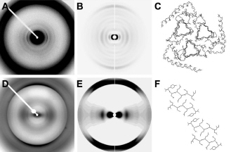
Diffraction from natural and recombinant prions: the observed x-ray diffraction pattern (A) from natural prions best fits the calculated pattern (B) from a 3-protofilament helical model (C); the observed x-ray diffraction pattern (D) from recombinant prions best fits the calculated pattern (E) from a stacked β-sheet model (F). (From H. Wille et al.,, Proc. Natl. Acad. Sci. USA 106(40), 16990 [2009], Copyright 2009 National Academy of Sciences, U.S.A.)

It is hard to believe that a single protein can be responsible for the damage inflicted by diseases such as human Creutzfeldt-Jakob and bovine spongiform encephalopathy (Mad Cow Disease). Yet the implicated protein, known as a prion and only about 200 amino acids long, can initiate and propagate a disease cycle just by changing its shape. Prions are amyloids, which are misfolded proteins now implicated in numerous diseases. Studying the prion diseases has required patience and fortitude because of disorder and insolubility of the prion samples. Aided by four U.S. Department of Energy x-ray beamlines (BioCARS 14-BM and BioCAT 18-ID at the Advanced Photon Source at Argonne, the 4-2 beamline at the Stanford Synchrotron Radiation Laboratory, and 12.3.1 at the Advanced Light Source), a …

more ...

A Closer Look at Protein Breathing

Fig. 1. A false-color scattering pattern from the protein myoglobin.

To take a static view of proteins and regard them as simple strings of amino acids that do grunt work in cells would be a mistake. Decades of biomedical research have proven that proteins are often large, complex in structure, and, as is becoming increasingly apparent, undergo sophisticated changes in space and time in order to keep cells functioning properly. Some proteins, when in solution, exhibit dramatic fluctuations in their three-dimensional structures, movement that looks like breathing. Because this movement has usually been studied in relatively dilute solutions, and not in the crowded interior of a cell, it has been difficult to know how much of the motion would actually occur in living systems. Recognizing the need for a new approach to the problem, researchers used the APS to study the breathing motions of a diverse group of five animal proteins. Their results provide badly needed modeling of protein movement in solution and data that can be used widely in biomedical applications, such as therapeutic drug design.

The researchers from Argonne National Laboratory and the Illinois Institute of Technology used computational modeling and wide-angle x-ray scattering (WAXS) experiments performed on …

more ...

Revealing the Structural Secrets of Plant Viruses

Fig. 1. Segments of the potyvirus soybean mosaic virus; several virions are also shown in cross-section. The symmetry and low-resolution structure of this virus are very similar to those of the potexvirus potato virus X, suggesting that flexible filamentous plant viruses share a common coat protein fold and approximate helical symmetry.

Viruses are extremely successful at finding ways to circumvent just about every host defense system. The secrets to this success seem to lie in their simplicity—small genomes and a metabolism that relies in part on the biochemistry of the host— and in their elegant, often breathtakingly beautiful and highly functional structures. Viral architecture, especially the coat protein structure, is intricately intertwined with successful invasion and infection of the host. Yet for many viruses, and particularly for a very large group of plant viruses, details of their structures have remained elusive. But researchers using a high-brilliance x-ray beamline at the APS have obtained important details about the structures of a soybean and a potato virus. This is good news for crop scientists concerned with finding ways to combat viral infestations.

Over the years, scientists have has invested much time and effort searching for methods to learn more about the …

more ...