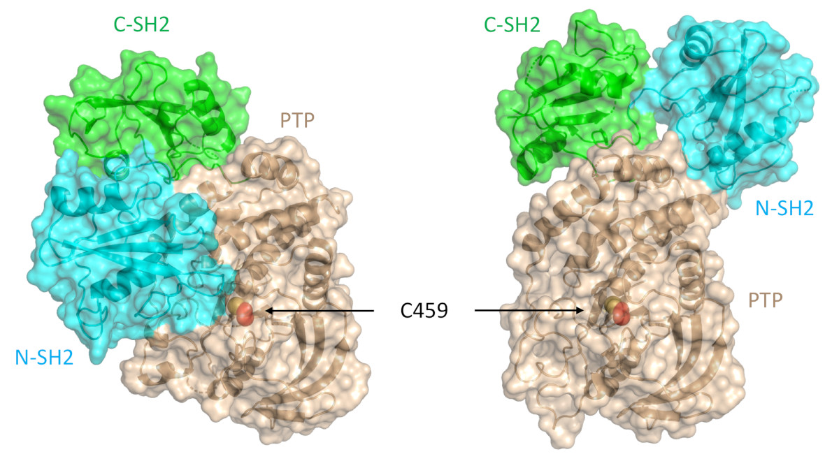
Structure-Function Understanding of aGPCR ECRs Critical for Drug-Design

Cellular communication mediated by a variety of cell-surface receptors involves ligand induced conformational changes in the extracellular region (ECR). A variety of drugs such as cetuximab (Epidermal Growth Factor Receptor), etrolizumab (Integrins), and erenumab (calcitonin receptor-like receptor) function by trapping ECRs in specific conformations and have proved to be effective therapeutic agents in several cancers, bowel diseases, and migraine. Leon et al., have addressed a class of relatively understudied G-protein couple receptors (GPCRs) called adhesion-GPCRs (aGPCRs) which have a structurally unique ECR with a diverse set of mechanistic possibilities. Specifically, they study the Gpr126, which is known to be essential for Schwann cell myelination, involved in heart development in the mouse model, and inner ear development in Zebra fish. Determination of the high-resolution structure of the Zebra fish Gpr126 ECR revealed the involvement of a heretofore undefined domain that has splice variants with or without a 23aa stretch in different isotypes (-ss or +ss) and is the primary determinant of whether the ECR is in the open active state or the closed inactive state. Also remarkable was the calcium dependent site at the tip of the ECR which promoted the closed inactive state of Gpr126. The closed conformation observed in …

more ...

Cool Temperatures During Hibernation May Freeze Muscle Contraction to Save Energy

Low temperatures shift myosin heads from an ordered relaxed state to a disordered state that cannot bind actin in response to stimulation.

Striated muscle contraction is a highly regulated process that involves an orchestrated series of events within the muscle’s contractile units, which are also known as sarcomeres. In a recent study, researchers studied the effect of low temperature on mammalian skeletal muscle contraction. They found that cooler temperatures reduce force generation by trapping filaments in the muscle sarcomeres in a refractory state that cannot undergo contraction and utilize adenosine triphosphate (ATP). This mechanism provides important insight into how hibernating animals may conserve energy while still allowing vital functions in the body to continue.

Muscle tissue’s repeating functional units, the sarcomeres, impart a striated or striped appearance to the tissue when examined under a microscope. Sarcomeres contain thin (containing the protein actin) and thick (containing the protein myosin) filaments that power contraction of skeletal muscle and cardiac muscle, both of which are striated.

Contraction of striated muscle is triggered when calcium enters the muscle fibers. This leads to structural changes in proteins known as troponin and tropomyosin that reside in the thin filament. Ultimately, this allows actin and …

more ...

Frustration and Folding of a TIM Barrel Protein

Multiple folding pathways discovered by simulations, validated against the time resolved SAXS and FRET data.

In their continuing endeavor to understand misfolding proteins as part of the etiology of a variety of diseases, the Matthews lab particularly focuses on the different factors that impede a protein’s path from the unfolded state to the global free energy minimum. The complexity of the folding trajectory understandably depends on the size of the protein mostly because of the formation of intermediates many of which often stall the formation of an optimal native conformation.

The triosphosphate isomerase (TIM) barrel family of proteins have very well characterized and conserved folding path despite large variations in sequence which make it an ideal group of proteins to obtain widely applicable insights into the folding process. Like other proteins studied before, S.solfataricus indole-3-glycerol phosphate synthase (SsIGPS) a TIM barrel protein goes through a burst phase followed by a relaxation phase and then eventually folds into the native conformation. Mutational and hydrogen exchange experiments have helped characterize the species found in the few millisecond time range of the folding process. In this study, Halloran et al., have used a combination of continuous flow FRET, SAXS and simulations …

more ...

Structure of BS Ric8A, a regulator of G-protein Biology

A model of the Ric8a protein structure showing the best scoring flexible tail (right), and the validation of the model against the SAXS data (left).

G-protein signaling has been the dominant theme in the Artemyev lab and their recent work specifically addresses Ric8A (Resistance to inhibitors of cholinesterase 8A) structure and function. Ric8A is a well-known regulator of G-protein biology and belongs to a class of proteins different from the G protein-coupled receptors (GPCRs), which act via interactions with monomeric Gα subunits as opposed to heterotrimeric Gαβγ proteins. SAXS was used in combination with crystallography and biochemical studies to show that the flexible C-terminal tail is important for the overall stability of Ric8A and the function as a guanine nucleotide exchange factor (GEF). The crystal structure revealed that Ric8A belongs to a functionally diverse class of proteins with what is known as an armadillo-fold (ARM) characterized by two layers of alpha helices arranged in a right handed superhelix. Ric8A diverged from the class in terms of the number of ARM repeats (8 as opposed to 10) and is further followed by a flexible region spanning ~ 70 residues.

Differential scanning fluorimetry (DSF) was used to determine that a construct without the …

more ...

Protein-Folding Mechanisms Elucidated Using Chaotic-Flow SAXS

The unfolded-state ensemble (USE) undergoes a continuous reduction in chain dimensions as a function of decreased denaturant concentration.

While there is emerging consensus in the protein folding community concerning the behavior of proteins under unfolding conditions, the occurrence of unfolded states under physiological (native) conditions and their propensity to aggregate are the basis of several human pathologies. Valuable insights into these transient species were obtained by taking advantage of the temporal resolution afforded by combining time-resolved fluorescence and continuous (in this case chaotic) flow SAXS (CF-SAXS) with all atom simulations and polymer theory. A group of researchers led by the Raleigh lab (Stony Brook University) used the 59 amino acid N-terminal domain of the ribosomal protein L9 (NTL9), which has a well-studied two state folding mechanism. By introducing FRET pairs several pairwise distance distributions were measured in the unfolded and native conditions in equilibrium and also the unfolded states in native conditions using a continuous flow mixer Interestingly chain contraction as indicated by fluorescence decay was observed well within the dead time of the mixer (~40 µs) showing that chain collapse happens considerably faster than the time-scale required for completion of the folding process (2.5 ms for NTL9 …

more ...

Probing the Powering of Contractions in Heart Failure

Sample x-ray diffraction patterns of resting cardiac muscle.

Current treatments can slow progression of heart failure, but do not address the underlying issues, including specific problems that cause systolic heart failure. In this condition, the heart doesn’t contract vigorously enough in pushing blood into the body’s circulation. But findings at nanometer and millisecond scales, based upon experimental data collected at BioCAT may help improve design of therapies directed at motor proteins to rescue failing hearts.

The heart muscle contractions that pump blood are generated by interactions between actin and myosin. These motor proteins power movement at the molecular level by converting the molecule ATP into energy. Earlier research in the lab of Michael Regnier, University of Washington (UW) professor of bioengineering, had shown that dATP, a natural variant of ATP, can be used to promote stronger heart function.

However, there remains a pressing need for data to explain why dATP helps to increase contractile force in heart disease. A new study headed by Regnier, who is a researcher at the UW Medicine Institute for Stem Cell and Regenerative Medicine and director of the Center for Translational Muscle Research, offers new insights, with unprecedented precision, about the nature of …

more ...

New Insights into Traumatic Brain Injury

X-ray diffraction patterns (a) and integrated data (intensity versus d-spacings) (b) of control and impact-loaded rat optic nerve samples.

Traumatic brain injury, or TBI, is often referred to as the “invisible injury” — while on the surface everything seems normal with brain structure, symptoms may present themselves in the behavior of the injured and cannot be explained. According to the Centers for Disease Control and Prevention, about 2.8 million TBI-related emergency department (ED) visits, hospitalizations and deaths occurred in the United States in 2013 alone. Every day, 153 people in the United States die from injuries that include TBI.

As reported in the Journal of Synchrotron Radiation, investigators from Joseph Orgel at the Illinois Institute of Technology, Argonne National Laboratory and the Army Research Lab studied the effect of controlled amounts of compressive force on rat optic nerves to attempt to identify the changes that occur in otherwise normal looking brain neurons due to the specific impact forces experienced during head trauma. This first-of-its-kind study used x-ray diffraction data obtained at the Biophysics Collaborative Access Team (Bio-CAT) 18ID beamline at the APS to examine the changes to myelin, the fatty material that wraps around nerve cell projections in …

more ...

A Target Mutation that Renders a Cancer Drug Ineffective

The wild-type enzyme SHP2 contains a protein tyrosine phosphatase catalytic domain (PTP domain) and two SH2 domains (N-SH2 and C-SH2). This version of SHP2 assumes a closed conformation in which the enzyme is autoinhibited. An allosteric inhibitor called SHP099 stabilizes SHP2. A specific mutation (E76K) to the gene, PTPN11, that encodes SHP2, results in a 120-degree pivot of the C-SH2 domain and relocalization of the N-SH2 domain. In this more open conformation of SHP2, the binding pocket for SHP099 is eliminated, thereby making SHP2 100 times more resistant to the allosteric inhibitor.

Mutations in the gene PTPN11, which encodes a common enzyme called SHP2, can result in developmental disorders, such as Noonan Syndrome, and act as oncogenic drivers in patients with certain blood cancers. Due to the well understood role of the enzyme SHP2 in Noonan Syndrome and in tumorigenesis, many companies are currently trying to develop drugs that inhibit the enzyme. However, it is unclear what impact mutations to SHP2 may have on the potential efficacy of drugs targeting this enzyme. Using the U.S. Department of Energy’s Advanced Photon Source (APS), researchers investigated the effect that the most frequently observed mutation of PTPN11 …

more ...

Unraveling the role of a “nebulous” protein

Force in muscle is generated by interactions between myosin motor proteins arranged in thick filaments and their binding sites on thin filaments made mostly of the protein actin. Nebulin is a giant actin-binding protein in skeletal muscle that is found along the length of the thin filaments but up to now, not much was known about what it does to affect muscle function. Mutations in the nebulin protein cause the muscles in patients with nemaline myopathy disease to be weak in patients, suggesting that properly functioning nebulin is important to generate force. This condition can be recapitulated in genetically engineered mice who have mutated nebulin or entirely lack nebulin so that the muscles in these animals also show weakness. Investigators from the University of Arizona and the Illinois Institute of Technology used the BioCAT beamline 18ID to study mice entirely lacking nebulin to discover the effects of nebulin on the nanoscale structure of muscle.

Using X-ray diffraction, they found that the thin filaments were found to be 3-fold less stiff in nebulin-deficient muscles and that the action of other proteins that normally function to turn on and off the thick filament were impaired. As a consequence, fewer myosin motors are …

more ...

This Boot was Made for Crosstalk

The first structure of a muntidomain human estrogen receptor alpha (hERα).

Estrogen receptor alpha (ERα) plays an important role in the development of various physiological functions; in particular, over 70% of breast cancers are ERα-positive. Although initial treatments that target ERα are often successful, many patients are not responsive or develop resistance so new treatment strategies are urgently needed. ERα is a nuclear hormone receptor that consists of a DNA-binding domain (DBD) and a ligand-binding domain (LBD) that binds the physiological hormone, estrogen, and functions as a hormone-regulated transcription factor. The structure of each domain is known but it is not clear how these two domains communicate with each other. Thus, there is no way to screen for drugs that disrupt this crosstalk. Now, work from a team of researchers from Case Western Reserve University has probed the structure of the complex and its domain interface. The team developed an elegant new approach, based on the new structure, to screen small molecules that target the bridging interface in ERα-positive breast cancer, with the potential for developing new pharmaceuticals to interrupt that communication.

The ERα project was the team’s first application of their iSPOT structural biology platform that combines …

more ...