A Super-relaxed Myosin State to Offset Hypertrophic Cardiomyopathy

Figure (a) shows a diffraction pattern from untreated muscle compared to treated muscle on the right. Intensification of the x-ray reflections from the treated muscle indicate a highly ordered “super-relaxed” state of myosin motors. Figure (b) shows the myosin heads in the compact “interacting head motif” which the heads adopt in the super-relaxed state allowing them to be packed closely and tightly on the surface of muscle thick filaments.

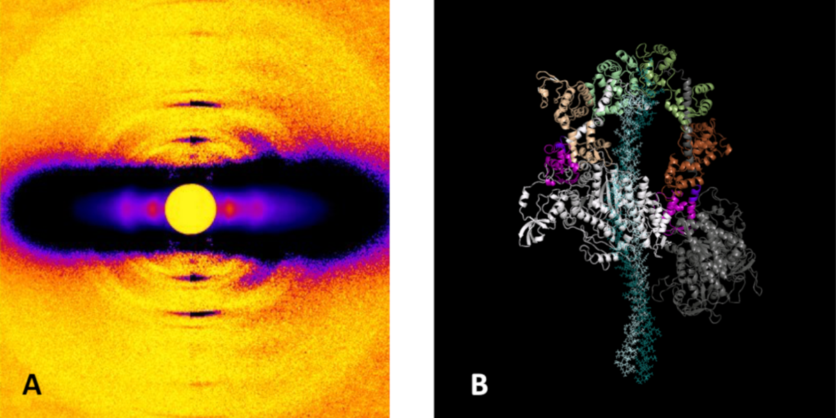
At its most basic level, the proper functioning of the heart depends upon the intricate interaction of proteins that trigger, maintain, and control the muscular contractions and relaxations of this vital organ. Disruption of those interactions can cause serious pathologies such as hypertrophic cardiomyopathy (HCM). Such disruptions can originate with mutations in the primary motor protein involved in heart contraction, ß-cardiac myosin, which can alter the rate of ATP hydrolysis and have been hypothesized to destabilize its super-relaxed state (SRX). Researchers investigated the stabilizing action of mavacamten, a cardiac drug currently in phase 3 clinical trials, on the ß-cardiac myosin super-relaxed state and its possible therapeutic effects on HCM. Their work, which included electron microscopy and low-angle x-ray diffraction imaging at the U.S. Department of Energy’s Advanced Photon Source …

more ...

Mechanistic Insights into Insulin Degrading Enzyme from Laminar-Flow SAXS

A model of IDE conformational states relevant to substrate capture and catalysis.

The Tang lab (University of Chicago, Illinois) has a deep interest in the structure and function of Insulin Degrading Enzyme (IDE), a zinc metalloprotease with a role in insulin and amyloid β degradation and known to be a significant factor in the pathophysiology of conditions such as Diabetes mellitus and Alzheimer’s disease. Previous structural studies showed that IDE forms dimers that can assume two distinct and functionally significant conformations. Substrate recognition occurs in an enclosed catalytic site formed by the dimerized IDE mostly via electrostatic interactions and size and shape complementarity. Catalytically important open conformations have been difficult to study using crystallography. The Tang lab used a combination of cryoEM, X-ray crystallography, Hydrogen-Deuterium exchange coupled Mass Spectrometry (HDX-MS), and SAXS to elucidate key mechanistic details. Four heretofore unknown and distinct conformations were determined using cryoEM strongly suggesting that substrate recognition and capture are contingent on the openness of IDE conformation.

Equilibrium SAXS showed that while IDE dimers exist in equilibrium between open and closed conformations, the presence of substrate conformationally restricts IDE to the partially closed or closed state. The question therefore was whether the enzymatic …

more ...

Shining X-ray Light on Heart Disease

Normal mouse hearts (WT) and hearts with a mutation causing Dilated Cardiomyopathy showing the effect of the mutation on heart structure.

Dilated cardiomyopathy (DCM) is a serious, and poorly understood, heart disease that affects about 1 million people in the United State. DCM is a progressive disease with no current cure, often culminating in heart transplantation. Many cases of DCM are caused by inherited gene mutations often located on specific muscle proteins that are part of the cell machinery that allows contractions. One such protein is Myosin Light Chain 2 (MYL2), part of the “motor” that powers contraction. The investigators identified a human mutation that causes DCM called D94A, and created a transgenic mouse that expresses this mutation allowing the mouse heart muscle to be studied by a wide range of techniques including X-ray diffraction at the BioCAT Beamline 18ID at the Advanced Photon Source, Argonne National Laboratory. The X-ray experiments showed that one of the reasons the muscle does not contract correctly is that the myosin ”motor” proteins are positioned further away from their targets than in normal heart muscle making it harder for them to generate the correct amounts of force to pump the right amount of blood …

more ...

Proteins May Prevent Dysfunction and Disease by Relaxing

Determining how proteins function on a molecular level is crucial to understanding the underlying basis for disease. Now scientists using the U.S. Department of Energy’s Advanced Photon Source (APS) at Argonne National Laboratory are one step closer to unraveling the mystery of how intrinsically disordered proteins work, according to new research published in Science. The team used simulations and high-brightness x-rays to tackle the question. Their results conclude that these proteins remain unfolded and expanded as they float loose in the cytoplasm of a cell.

Proteins are chains of amino acids that fold into three-dimensional structures, giving them their shape and determining the way they interact with other molecules. Many proteins form rigid structures, but intrinsically disordered proteins (IDPs) are “floppy” and do not fold into a regular structure. These disordered proteins are floppy because their parts interact just as well with water as with each other. Up to 30 percent of all proteins are disordered — and must be disordered in order to work properly.

Researchers have struggled to understand precisely how disordered IDPs are — and how they work. Their floppy structures make it difficult to extract their exact dimensions, making the extent of that disorder, along with …

more ...

The Chd1 Chromatin Remodeler Shifts Nucleosomal DNA Bidirectionally as a Monomer

DNA in eukaryotic cells is normally found associated with protiens called histone in particles called nucleosomes linked by short segments of DNA. Chromatin remodelers are specialized ATP-dependent molecular machines that can reorganize the structure of nucelsomes as needed for such processes such as replication, transcription and DNA repair.

Remodeling results in evenly spaced nucleosomes along the DNA strand yet the way remodelers achieve this is not understood. CHD-1 is a remodeler important for transcription. Here, the authors show that the Chd1 remodeler shifts DNA back and forth by dynamically alternating between different segments of the nucleosome. During sliding, Chd1 generates unstable remodeling intermediates that spontaneously relax to a pre-remodeled position. They demonstrate that nucleosome sliding is tightly controlled by two regulatory domains: the DNA binding domain, which interferes with sliding when its range is limited by a truncated linking segment, and the chromodomains, which play a key role in substrate discrimination. They propose that active interplay of the ATPase motor with the regulatory domains may promote dynamic nucleosome structures uniquely suited for histone exchange and chromatin reorganization during transcription. This work advances our understanding of the Chd1 chromatin remodeler, and puts forward several concepts that may also apply …

more ...

Perplexing cooperative folding and stability of a low-sequence complexity, polyproline 2 protein lacking a hydrophobic core

The physical basis of protein folding stability and cooperativity remains a topic of great interest. Folding of globular proteins is generally assumed to be driven by energetically favorable burial of hydrophobic groups and that early development of secondary structure increases the cooperativity of folding. The Sosnick group at the University of Chicago examines these assumptions in a protein (snow flea antifreeze protein (sfAFP) that is striking in its dearth of hydrophobic burial and its lack of α and β structures, while having a low sequence complexity with 46% glycine. The interior of the protein is filled only with backbone H-bonds between six polyproline 2 (PP2) helices. Unexpectedly, the protein folds in a kinetically two-state manner and is moderately stable at room temperature, similar behavior to that observed for typical globular proteins having α and β structures and a hydrophobic core. Hence, these features are not necessary for folding cooperativity and stability. This enigma forces a reexamination of the possible combination of factors that can stabilize a protein. The authors propose that a major part of the stability arises from the unusual match between residue-level PP2 dihedral angle bias in the unfolded state and PP2 helical structure in the native state …

more ...

Stress-Triggered Phase Separation Is an Adaptive, Evolutionarily Tuned Response

In eukaryotic cells, diverse stresses trigger coalescence of various RNA binding proteins into so-called stress granules. In vitro, stress-granule associated proteins have been observed to demix to form liquids, hydrogels, and other assemblies. Demixing of an abundant RNA-binding protein into hydrogel droplets, triggered by stress-associated physiological conditions, appears to promote cell fitness during stress.

Here, a U Chicago based team lead by D. Allan Drummond and Tobin Sosnick showed that poly(A)-binding protein (Pab1 in yeast), a defining marker of stress granules, phase separates and forms hydrogels in vitro upon exposure to physiological stress conditions. Other RNA-binding proteins depend upon low-complexity regions (LCRs) or RNA for phase separation. Based on unique evolutionary patterns, the authors created LCR mutations, which systematically tune its biophysical properties and Pab1 phase separation in vitro and in vivo. Mutations that impede phase separation reduced organism fitness during prolonged stress. Such mutations reduce the thermal and pH sensitivity of Pab1’s demixing and, hence, reduce fitness during growth at high temperature and during energy depletion, indicating that demixing is adaptive. Together, the results illuminate a uniquely complete path from evolved sequence features, to phase separation, to stress-triggered demixing, and finally to organism fitness …

more ...

Asymmetric unwrapping of nucleosomal DNA propagates asymmetric opening and dissociation of the histone core

Nucleosomes are protein–DNA structures which eukaryotic organisms use to package and organize DNA inside the nucleus. Nucleosomes need to be disassembled to permit transcription of DNA and reassembled afterwards, hence are an important component of the gene regulation machinery. The nucleosome core particle (NCP) consists of DNA wound around a core of eight histone proteins including two dimers of H2AH2B histones and an (H3–H4) tetramer that is assembled as a dimer of dimers. A team led by Lois Pollack at Cornell University used time-resolved SAXS at the BioCAT beamline 18ID at Advanced Photon Source and TR-FRET, in collaboration with Lisa Gloss, at Washington State University to study changes in the DNA conformations as a function of the composition of the histone core during salt induced disassembly of NCPs in order to allow identification of kinetic pathways and transient intermediates that show how the sequence of events involving DNA unwrapping and protein dissociation are connected. The investigators found that H2AH2B histone dimers are released sequentially with an octasome-to-hexasome transition guided by asymmetric unwrapping of the DNA. This work suggests a mechanism for NCP remodeling in which DNA conformation facilitates the reconfiguration of the histone core. This …

more ...

The POTRA domains of Toc75 exhibit chaperone-like function to facilitate import into Chloroplasts

Chloroplasts, like mitochondria, are organelles of endosymbiotic origin, having evolved from initial engulfment of a cyanobacterium by a eukaryotic cell. Most of the bacterial genome was subsequently lost so that most proteins found within chloroplasts are synthesized in the cytoplasm as preproteins and then imported via specialized machinery prior to trafficking to their final destination. Protein import is accomplished by the TOC (translocon on the outer chloroplast membrane) and TIC (translocon on the inner chloroplast membrane) machineries in the outer and inner envelope membranes, respectively. The TOC complex includes a protein called Toc75, which serves as the translocation channel along with two other proteins, as well as Toc33 and Toc159, which both contain GTPase domains, which help drivesubstrate selection and importation. Structural information for the TOC complex was hitherto lacking, hindering the ability of investigators to form mechanistic models for function. Here a team lead by Nicholas Noinaj (Purdue University) and Danny Schnell (Michigan State University) reported crystals structures of Toc75 consisting of three tandem POTRA domains. High quality size exclusion chromatography coupled SAXS experiments at the BioCAT Beamline 18ID were important in establishing that crystals structures accurately represented protein structure in solution. The POTRA domains may help facilitate preprotein …

more ...

Solution Structure of the HIV-1 Intron Splicing Silencer and Its Interactions with the UP1 Domain of Heterogeneous Nuclear Ribonucleoprotein (hnRNP) A1

Human immunodeficiency virus type 1 (HIV-1) requires controlled synthesis of its protein complement for persistent infection and successful virion production. Genome expression is tightly regulated at the levels of transcription, splicing, mRNA nuclear export, and translation. RNA polymerase II-dependent transcription yields a 9-kilobase (kb) polycistronic transcript that undergoes multiple rounds of alternative splicing to produce upward of 100 different viral mRNAs that recruit antagonistic host RNA-binding proteins. Thus, HIV-1 splicing pathways are essential components of the viral replication cycle and represent new targets for therapeutic intervention. Because HIV-1 splicing depends on protein-RNA interactions, it is important to know the tertiary structures surrounding the splice sites. Herein, we present the NMR solution structure of the phylogenetically conserved ISS stem loop. ISS adopts a stable structure consisting of conserved UG wobble pairs, a folded 2X2 (GU/UA) internal loop, a UU bulge, and a flexible AGUGA apical loop. SEC-SAXS data collected at BioCAT was used to confirm the general features of the NMR structure and by refining the NMR structure against the SAXS data, obtain a better structure than that of NRM alone. Collectively, this work provides additional insights into how HIV-1 uses a …

more ...