Storing the Power to Fly

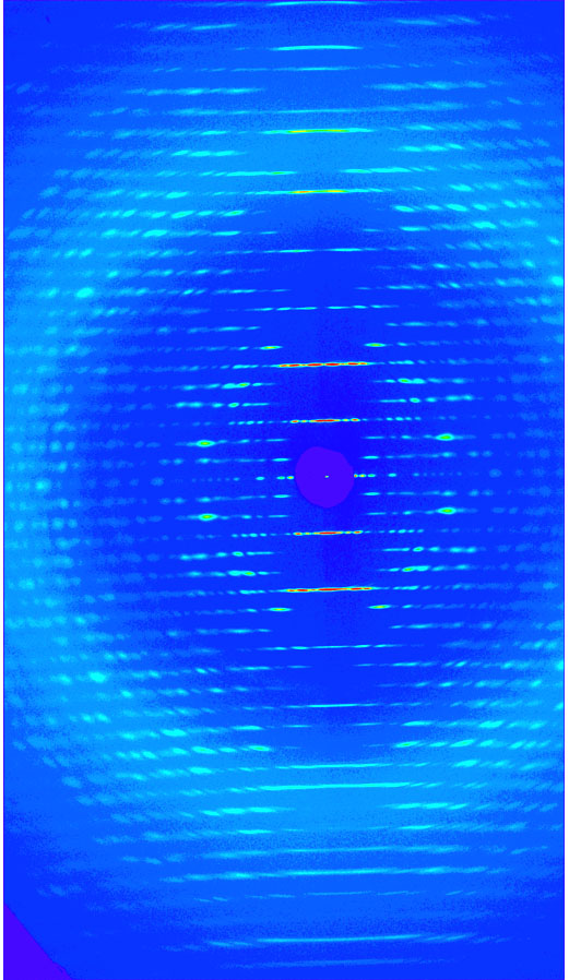
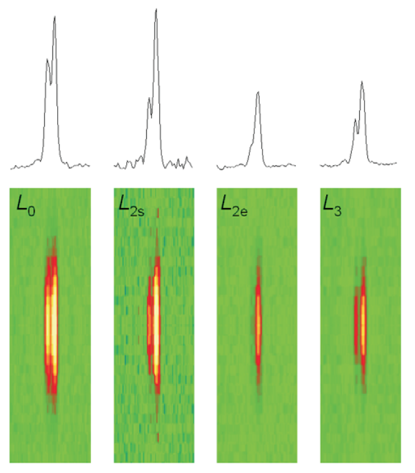
Fruit flies beat their wings faster than their cellular powerplants can generate the energy needed for flapping. To resolve this energetic discrepancy, researchers from the California Institute of Technology, the Illinois Institute of Technology, and the University of Vermont used the BioCAT beamline 18-ID at the APS to obtain a series of x-ray photographs that revealed the flies’ secret: A muscle protein used to power wings acts like a spring, storing energy while stretched before snapping back. Not only did this finding surprise researchers who study muscle, but the results might also help scientists better understand the human heart.

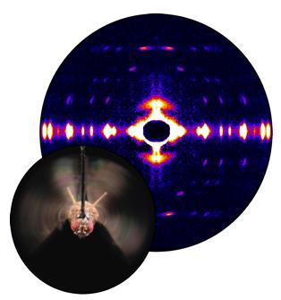
Drosophila, the fruit fly, beats its wings up and down once every 5 milliseconds. Two layers of muscle control this action: when one layer contracts, it stretches the other. The stretched muscle is then primed to contract; when it does, it stretches the first layer and completes the cycle. These cycles occur faster than nerve impulses can stimulate them; hence, the muscles themselves must keep the beat going.

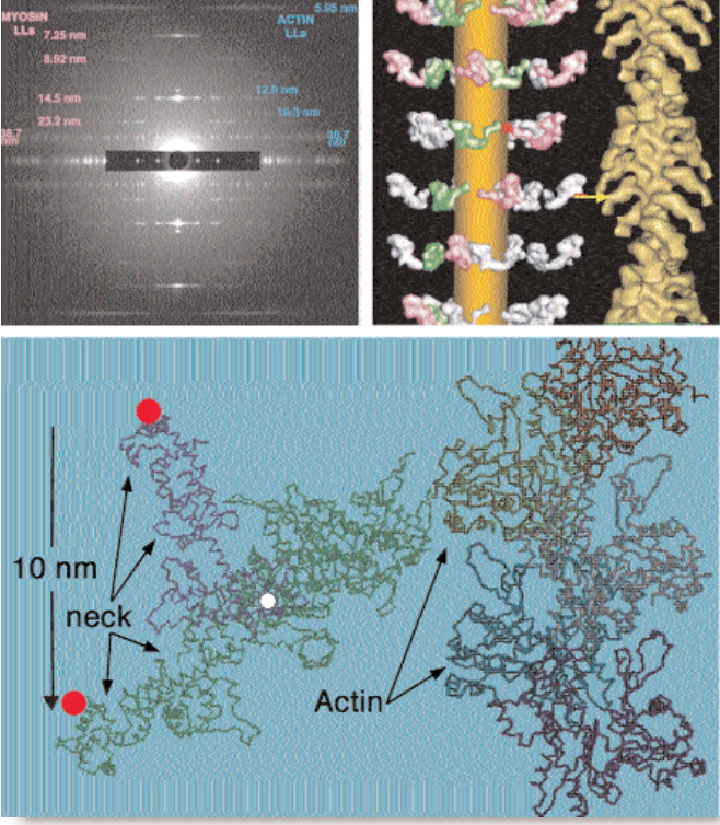
To find out how muscles do this, the research team needed to visualize the proteins that make muscle contract. Within the flight muscle cells, two proteins cooperate to do this. Myosin proteins …

more ...

Atomic Models of Plant Viruses

Amy Kendall (left), Michele McDonald, and Sarah Tiggelaar — all with the Stubbs Lab in the Department of Biological Sciences at Vanderbilt University — taking data at the BioCAT 18-ID beamline.

Kendall: “Our lab uses fiber diffraction data from oriented sols and dried fibers to determine the structures of flexible filamentous plant viruses. Our long-term goal is to produce atomic models based on diffraction data at resolutions close to 3 Å, but a great deal of information can be obtained at lower resolution, including the size, shape, and symmetry of the viruses.

These viruses are of great significance as models for fundamental virology and cell biology, They have enormous potential as vectors in biotechnology and they are of considerable importance because of the damage they cause to agriculture. We also use the filamentous plant viruses as important model systems for developments in fiber diffraction.”

more ...

Measuring the Efficiency of the Myosin Motor at High Load

Fig. 1. Two-dimensional (2-D) patterns in the region of the M3 reflection recorded on a charge-coupled device detector at the Bio-CAT beamline with 3-m camera-to-sample distance, normalized by X-ray exposure time. Total exposure times: L0, 25 ms; L2s, 5.5 ms; L2e, 21 ms; and L3, 25 ms. Fiber cross-sectional area equals 38•103 μm2; isometric tension equals 230 kPa; sarcomere length equals 2.16 μm. On the top are shown the intensity profiles obtained by integrating 80 pixels on each side of the meridional axis of the 2-D patterns.

The sliding filament model of muscle contraction is more than 50 years old, yet theories about the precise mechanisms of the motor function still generate some controversy. A group of researchers from Università di Firenze, Istituto Nazionale di Fisica della Materia, Brandeis University, King’s College London, the European Synchrotron Radiation Facility, Illinois Institute of Technology, and Argonne National Laboratory used insertion device beamlines at BioCAT at the APS and at the European Synchrotron Radiation Facility (ESRF) to take a closer look at the molecular structure of myosin II, the molecular motor in muscle, as it works under different loads. This study provides …

more ...

Finding Active Proteins

When combinatorial chemistry produces new varieties of reagents, the tricky next step is figuring out whether those molecules will be biochemically active. While there are several methods for finding active molecules, they all have limitations. Researchers from Argonne National Laboratory, using the BioCAT beamline 18-ID, employed wide-angle x-ray scattering (WAXS) to develop a method for identifying drug candidates.

One way to identify active molecules is to design an assay for the desired function of a protein target and then test each small molecule variant. This approach is labor intensive and time consuming because it means that a different assay must be developed for each drug target of interest. Another approach is based on deriving a more general probe that would identify whether any protein structural change had occurred. A large number of samples could be screened in this way; those that showed promise could then be examined more closely for mechanistic details. Just such a general method, which uses x-ray scattering to detect functional drug candidates, is now available.

The APS served as a unique resource to aid the investigators in their study of WAXS of proteins in solution. The researchers are developing WAXS as a probe for detecting …

more ...

How Water Molecules are Connected

Water may be the most important molecule on Earth, but our understanding of its properties is embarrassingly limited. In solid ice form, water takes on numerous phases and structures that can be studied by means of diffraction techniques. As a liquid, however, water poses a frustrating structural puzzle because of the complex hydrogen bonding that forms a disordered network. Recently, researchers from the Stanford Synchrotron Radiation Laboratory, the BESSY laboratory, Stockholm University, Linköping University, and Utrecht University have used the BioCAT 18-ID beamline at the APS, as well an Advanced Light Source (ALS) beamline, to obtain detailed information about the nearest neighbor coordination geometry in liquid water.

Previous experimental efforts to understand water structure have relied on several methods including infrared spectroscopy and neutron and x-ray diffraction. Unfortunately, the structural information provided by infrared spectra is ambiguous for water, and diffraction provides only radial distribution functions that do not allow unique assignment of local hydrogen bonding configurations. In the work reported by Wernet et al., x-ray absorption spectroscopy (XAS) and x-ray Raman spectroscopy (XRS) were used to investigate local bonding in the first coordination shell of water. In XAS, x-rays are absorbed by core electrons close to the nucleus …

more ...

Grasping the Structure of Insect Muscle Poised to Contract

Fig. 1. Top left: Low-angle x-ray diffraction pattern from relaxed insect flight muscle. (Pink and blue numbers represent spacings between repeating structural elements as calculated from the pattern.) Top right: Structure (left) of the modeled myosin heads (pink/white/green) in their relaxed, pre-powerstroke state and (right) in their “rigor” or post-powerstroke state. Bottom: Transition from the head shape in relaxed insect muscle (blue) to rigor-like final shape (green), showing the pivots needed (red on thick filament, white within head) to let the neck swing from one form to the other when bound to actin.

Researchers have achieved the first detailed view of resting muscle filaments poised to contract, a long sought window into the biochemical cycle that causes muscle contraction. The group determined the overall structure of insect muscle fibers from x-ray diffraction patterns and performed computer modeling to analyze the data. The resulting structure, reported in August 2003, suggests a specific mechanism for insect muscle activation.

Muscle cells (fibers) contain two parallel, overlapping sets of protein filaments, made of myosin and actin, respectively, that align with the direction of contraction. The myosin and actin filaments have fixed length individually, but they slide past each other like telescope segments …

more ...

Role of Interfilament Spacing in Muscle Filament Calcium Sensitivity

The heart regulates ventricular output in response to changes in ventricular filling, a mechanism known as Frank Starling’s law of the heart. Part of the cellular basis for the law is an increase in myofilament Ca2+ responsiveness upon an increase in sarcomere length (SL). (Sarcomeres are the basic functional units of muscle fibers, which contract in response to calcium influx.) How information on SL is transmitted to myofilaments is unclear. One prominent hypothesis is that a decrease in myofilament lattice spacing (LS) in response to an increase in SL underlies the Ca2+ sensitivity.

In support of this hypothesis, shrinkage of the myofilament lattice by osmotic compression using high-molecular-weight moieties has been shown to increase myofilament Ca2+ sensitivity at a short SL, mimicking the effect of increased SL. The extent of LS shrinkage in that experiment was estimated from the reduction in muscle width, however. Recently, researchers directly measured LS by synchrotron x-ray diffraction as a function of SL in skinned rat cardiac trabeculae bathed in 0-6% dextran (high-molecular-weight) solutions.

To assess the effect of SL on myofilament Ca2+ sensitivity, Ca2+-contraction force relationships were determined at three SLs (1.95, 2.10, and 2 …

more ...

The “Second Stalk” of ATP Synthase: Dimerization Domain Structure

Fig. 1. Function and structure of the b2 dimerization domain in ATP synthase. ATP synthesis is catalyzed as follows. Proton flow through the membrane-embedded F0 portion causes the ring of c subunits and the γε “central stalk” to rotate, which bridges the F0 and F1 complexes. As the central stalk rotates, it drives conformational changes in the β-subunits. The “second stalk” of the complex is composed of the b2 and δ subunits. Its function is to provide a stator to prevent rotation of F1. A model for the dimerization domain of b2, derived from the crystal structure of the b62-122 monomer and SAXS of the solution dimer, is shown to the right of the ATP synthase holoenzyme. The alanines that define the undecad repeat, characteristic of a right-handed coiled coil, are in green.

Hydrolysis of adenosine triphosphate (ATP) drives many of the vast range of energy-consuming processes within a cell. The ATP synthase enzyme is a molecular motor that couples proton movement to ATP synthesis. In the rotational model of the enzyme, protons flow down an electrochemical gradient, through the membrane-embedded F0 portion of ATP synthase, causing a ring of c …

more ...